Dog Joint Health And Arthritis Signs
Learn about dog joint health and arthritis signs with expert tips and data-backed advice.
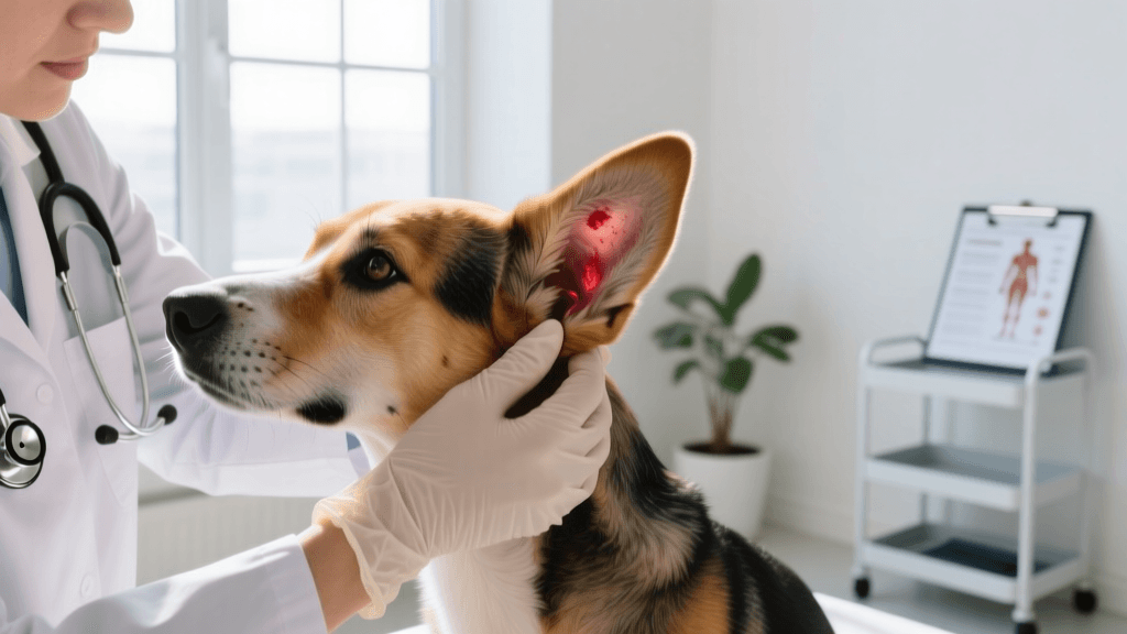
Recognising the Early Signs of Joint Problems in Dogs
Joint disease is one of the most common health conditions affecting dogs in the United Kingdom, yet it frequently goes undetected until significant damage has already occurred. The British Small Animal Veterinary Association (BSAVA, 2022) estimates that approximately 20% of dogs over the age of one year show some degree of joint disease, with that figure rising to over 80% in dogs aged ten years and older. Because dogs are instinctively inclined to mask discomfort, owners often mistake the early signs of arthritis for normal ageing, delaying treatment that could meaningfully improve quality of life.
Understanding what to look for — and when to act — is the single most important thing a dog owner can do to protect their pet's long-term mobility. Joint health is not simply a concern for elderly dogs; large and giant breeds such as Labrador Retrievers, German Shepherds, and Rottweilers can begin showing degenerative changes as early as two to three years of age, particularly if they have a history of developmental conditions like hip dysplasia or osteochondrosis.
What Is Canine Osteoarthritis?
Osteoarthritis (OA) in dogs is a progressive, degenerative joint disease characterised by the breakdown of articular cartilage — the smooth tissue that cushions the ends of bones within a joint. As cartilage erodes, the underlying bone becomes exposed, leading to inflammation, pain, and the formation of bony outgrowths called osteophytes. Over time, the joint capsule thickens, the surrounding muscles weaken from disuse, and the dog's range of motion becomes increasingly restricted.
The condition is classified as either primary or secondary. Primary OA arises without a clear preceding cause and is most often linked to age-related wear. Secondary OA, which is far more common in dogs, develops as a consequence of another condition — cruciate ligament rupture, hip or elbow dysplasia, patellar luxation, or previous joint trauma. According to the People's Dispensary for Sick Animals (PDSA, 2023), cruciate ligament disease alone accounts for a significant proportion of OA cases in dogs under seven years of age, particularly in Labrador Retrievers and Staffordshire Bull Terriers.
Joints Most Commonly Affected
While OA can affect any synovial joint in the body, certain joints bear disproportionate loads and are therefore more vulnerable. The hips, elbows, stifles (knees), and lower spine are the most frequently involved sites in dogs. Elbow dysplasia is particularly prevalent in large breeds, with studies conducted at the Royal Veterinary College (RVC) in London indicating that up to 17% of Labrador Retrievers carry some form of elbow pathology. Hip dysplasia, meanwhile, affects an estimated 15–20% of large-breed dogs in the UK, making it one of the most significant orthopaedic concerns in veterinary practice.
Behavioural and Physical Signs to Watch For
Because dogs cannot verbalise pain, owners must learn to interpret subtle changes in behaviour and movement. The signs of joint disease exist on a spectrum — from barely perceptible stiffness after rest to obvious lameness and reluctance to bear weight. Catching changes at the mild end of that spectrum gives veterinary teams the best opportunity to slow disease progression.
Common early indicators include a slight hesitation before rising from a lying position, reduced enthusiasm for walks that the dog previously enjoyed, and a tendency to lag behind on outings rather than pulling ahead. Some dogs begin to show irritability when touched around the affected joint, or they may lick, chew, or bite at the area persistently. Changes in posture — such as a hunched back or a shifted weight distribution — can also signal discomfort that the dog is attempting to compensate for.
- Stiffness or limping that is worse after rest and improves with gentle movement
- Reluctance to climb stairs, jump into the car, or get onto furniture
- Visible swelling or heat around one or more joints
- Muscle wasting (atrophy) in the hindquarters or around a specific limb
- Audible clicking or grinding sounds (crepitus) during movement
- Changes in temperament — increased irritability, withdrawal, or reduced interaction
- Altered gait, including a bunny-hopping motion in the hindlimbs
The Difference Between Stiffness and Lameness
Stiffness and lameness are related but distinct presentations. Stiffness typically refers to reduced ease of movement, particularly after periods of inactivity, and often resolves within a few minutes of gentle exercise as the joint warms up and synovial fluid redistributes. Lameness, by contrast, involves an observable alteration in weight-bearing — the dog actively avoids loading the affected limb, resulting in an uneven gait. Persistent lameness lasting more than 24 to 48 hours warrants a veterinary assessment, as does any sudden-onset severe lameness, which may indicate an acute injury rather than chronic degeneration.
Veterinary Diagnosis and Assessment Tools
A thorough orthopaedic examination is the foundation of any arthritis diagnosis. Your veterinarian will observe the dog's gait, assess posture and muscle symmetry, and systematically palpate each joint to identify pain responses, swelling, reduced range of motion, or crepitus. Gait analysis may be performed in the consulting room or, in specialist centres, using force plate technology that objectively measures weight distribution across all four limbs.
Radiography (X-rays) remains the most widely used imaging modality for confirming OA. Characteristic findings include joint space narrowing, subchondral bone sclerosis, osteophyte formation, and soft tissue swelling. It is worth noting that radiographic changes do not always correlate directly with the degree of pain a dog experiences — some dogs with severe radiographic OA show relatively mild clinical signs, while others with modest changes may be significantly uncomfortable. Advanced imaging such as CT scanning or MRI, available at referral centres including the Animal Health Trust in Newmarket and the Queen Mother Hospital for Animals at the RVC, provides greater detail for complex cases or pre-surgical planning.
Pain scoring tools, such as the Liverpool Osteoarthritis in Dogs (LOAD) questionnaire and the Canine Brief Pain Inventory (CBPI), allow owners to systematically record their observations at home and provide veterinarians with a more complete picture of how the condition affects daily life. These validated instruments are increasingly used in both primary practice and specialist settings to monitor treatment response over time.
Blood Tests and Ruling Out Other Conditions
While there is no blood test that directly diagnoses OA, haematology and biochemistry panels are often recommended before initiating long-term medication. Non-steroidal anti-inflammatory drugs (NSAIDs), which form the cornerstone of medical management, require healthy kidney and liver function for safe use. Baseline bloodwork establishes a reference point and identifies any pre-existing organ compromise that might influence prescribing decisions. In older dogs or those with concurrent health conditions, these tests should be repeated every six to twelve months during NSAID therapy.
Medical Management and Dosage Guidance
The management of canine OA is multimodal, meaning that the best outcomes are achieved by combining several approaches rather than relying on any single treatment. Pharmacological management, weight control, physiotherapy, and environmental modification each contribute to reducing pain and preserving function.
NSAIDs are the most commonly prescribed class of drug for OA pain in dogs. Licensed veterinary NSAIDs include meloxicam, carprofen, grapiprant, and robenacoxib, among others. Meloxicam is frequently used at an initial dose of 0.2 mg/kg on the first day, followed by a maintenance dose of 0.1 mg/kg once daily, administered with food to reduce the risk of gastrointestinal upset. Carprofen is typically dosed at 4 mg/kg once daily or 2 mg/kg twice daily. These are general guidelines; your veterinarian will determine the appropriate drug and dose for your individual dog based on body weight, concurrent conditions, and response to treatment.
"Multimodal analgesia — combining an NSAID with adjunctive therapies such as gabapentin, physiotherapy, and weight management — consistently produces better outcomes than any single intervention alone. Owners who engage actively with the full treatment plan report significantly higher satisfaction and observe greater improvements in their dog's mobility and demeanour."
— BSAVA Manual of Canine and Feline Musculoskeletal Disorders, 2nd Edition (BSAVA, 2022)
For dogs that cannot tolerate NSAIDs, or where additional pain control is needed, gabapentin is increasingly used as an adjunct. Typical doses range from 5 to 10 mg/kg two to three times daily, though this should always be prescribed and monitored by a veterinarian. Amantadine, an NMDA receptor antagonist, may be added in cases of central sensitisation at doses of approximately 3–5 mg/kg once daily. Injectable treatments such as bedinvetmab (Librela), a monoclonal antibody targeting nerve growth factor, represent a newer class of therapy that has shown promising results in clinical trials, with monthly subcutaneous injections providing sustained pain relief in dogs with moderate to severe OA.
Nutraceuticals, Joint Supplements, and Their Evidence Base
The market for joint supplements in dogs is substantial, and the quality of evidence supporting individual products varies considerably. Omega-3 fatty acids, particularly eicosapentaenoic acid (EPA) and docosahexaenoic acid (DHA) derived from marine sources, have the strongest evidence base among nutraceuticals. Studies suggest that supplementation at doses of 75–100 mg/kg of combined EPA and DHA per day can reduce inflammatory mediators within joint tissue and may allow a modest reduction in NSAID dose in some patients.
Glucosamine and chondroitin sulphate are widely used, though clinical trial results in dogs have been mixed. The PDSA (2023) notes that while these compounds are generally safe and well-tolerated, owners should not expect them to replace veterinary-prescribed analgesia in dogs with established OA. Green-lipped mussel extract, which contains a unique combination of omega-3 fatty acids and glycosaminoglycans, has shown some positive results in small-scale studies and is considered a reasonable adjunct by many veterinary practitioners.
| Supplement | Typical Daily Dose | Evidence Level | Notes |
|---|---|---|---|
| Omega-3 (EPA/DHA) | 75–100 mg/kg combined | Moderate–Good | Use marine-sourced products; monitor for GI effects |
| Glucosamine HCl | 20 mg/kg once daily | Limited | Safe; may support cartilage matrix |
| Chondroitin sulphate | 15 mg/kg once daily | Limited | Often combined with glucosamine |
| Green-lipped mussel | Varies by product | Emerging | Follow manufacturer guidance; whole-extract preferred |
| Boswellia serrata | Varies by product | Limited | Some anti-inflammatory properties reported |
Weight Management and Exercise as Therapeutic Tools
Body weight is one of the most modifiable risk factors for both the development and progression of OA. Every kilogram of excess body weight places approximately five additional kilograms of force through the hip and stifle joints during normal locomotion. Research from the University of Liverpool's veterinary school has demonstrated that dogs maintained at an ideal body condition score (BCS) of 4–5 out of 9 develop OA at a significantly later age and with less severity than overweight counterparts. In dogs already diagnosed with OA, even a 6–8% reduction in body weight has been associated with measurable improvements in mobility and pain scores.
Exercise prescription for arthritic dogs requires careful calibration. The goal is to maintain muscle mass and joint range of motion without exacerbating inflammation. Short, frequent walks on even surfaces are preferable to long, infrequent outings. Hydrotherapy — either underwater treadmill or swimming — is particularly valuable because the buoyancy of water reduces joint loading while allowing full muscle engagement. Many veterinary physiotherapy centres across the UK, including those affiliated with the Canine Hydrotherapy Association, offer structured rehabilitation programmes tailored to individual patients.
- Aim for two to three short walks daily (10–15 minutes each) rather than one long walk
- Avoid high-impact activities such as ball chasing, jumping, and rough play during flare-ups
- Introduce hydrotherapy sessions once or twice weekly if available and tolerated
- Incorporate passive range-of-motion exercises as directed by a veterinary physiotherapist
- Monitor the dog for 24 hours after any new activity — increased stiffness the following day suggests the session was too demanding
Environmental modifications at home can make a significant difference to daily comfort. Orthopaedic memory foam beds reduce pressure on painful joints during rest. Ramps or steps allow dogs to access furniture or vehicles without jumping. Non-slip mats on hard floors prevent the stumbling and compensatory muscle strain that smooth surfaces cause. Raised food and water bowls reduce the need for neck and shoulder flexion during feeding, which is particularly helpful for dogs with cervical or forelimb joint disease.
Regular veterinary reassessment — ideally every three to six months for dogs on long-term medication — allows treatment plans to be adjusted as the disease progresses or as the dog's response to therapy evolves. Open communication between owner and veterinary team, supported by structured pain scoring at home, remains the most reliable way to ensure that a dog with joint disease continues to live as comfortably and actively as possible.
Tom Renshaw
All our authors care for dogs every day — read more of their work on the authors page.