Cherry Eye in Dogs: Symptoms, Treatment Costs & Recovery Guide
Complete guide to cherry eye in dogs: symptoms checklist, surgical vs medical treatment options, costs ($300-$2,000+), breed risks, and recovery timeline.
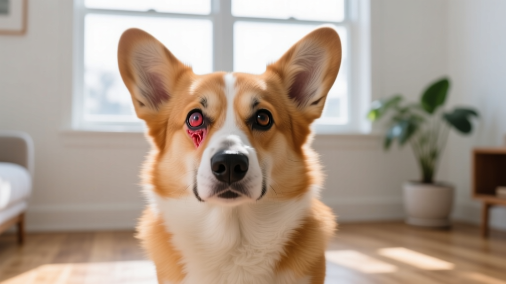
That bright red, fleshy mass suddenly appearing in the corner of your dog's eye can be alarming. Known as cherry eye, this condition occurs when the nictitating membrane gland (third eyelid gland) prolapses — meaning it pops out of its normal position and becomes visible as a pink or red bulge. According to the Merck Veterinary Manual, the third eyelid gland produces approximately 30-50% of a dog's aqueous tear film, making its proper function critical for long-term eye health.
Cherry eye is most commonly seen in young dogs under two years of age, with certain brachycephalic and sporting breeds disproportionately affected. While not immediately life-threatening, untreated cherry eye can lead to chronic dry eye (keratoconjunctivitis sicca), corneal ulceration, and permanent vision impairment. Early veterinary evaluation is essential — always consult your veterinarian for a proper diagnosis and treatment plan tailored to your dog's specific case.
What Is Cherry Eye in Dogs?
Cherry eye — medically termed prolapse of the nictitating membrane gland — happens when the connective tissue anchoring the third eyelid gland weakens or fails, allowing the gland to flip outward and protrude above the eyelid margin. The exposed gland becomes inflamed, swollen, and irritated due to environmental exposure and the dog's own pawing or rubbing.
The condition can affect one eye (unilateral) or both eyes (bilateral). When bilateral, the second eye may prolapse weeks or even months after the first. The exact cause remains partially idiopathic (unknown origin), but veterinary ophthalmologists believe a genetic weakness in the retinaculum — the fibrous tissue that secures the gland to the orbital rim — is the primary underlying factor.
Cherry Eye Symptoms: Early Warning Signs vs. Advanced Stages
Recognizing cherry eye early improves treatment outcomes and reduces the risk of secondary complications. Use this checklist to assess your dog's condition:
| Severity | Symptoms | Action Required |
|---|---|---|
| Early Stage | Small pink/red mass in inner corner of one eye; intermittent appearance (comes and goes); mild squinting; occasional pawing at eye | Schedule vet appointment within 1-2 weeks |
| Moderate Stage | Persistent prolapsed gland visible at all times; noticeable swelling and redness; increased tear production or mucoid discharge; dog frequently rubs face on furniture | Vet visit within 3-5 days |
| Advanced Stage | Gland appears dark red or purplish; thick mucopurulent discharge; corneal cloudiness or ulceration; dog keeps eye partially or fully closed; secondary conjunctivitis | Urgent vet visit within 24-48 hours |
| Complicated | Corneal ulcer (visible scratch or pit on eye surface); severe squinting and light sensitivity; blood in tear film; eye appears sunken or shrunken; bilateral involvement with both glands prolapsed | Emergency veterinary or veterinary ophthalmologist immediately |
The intermittent nature of early cherry eye catches many owners off guard. You may notice the gland pop out during car rides, after naps, or when your dog is excited — then seemingly disappear. This does not mean the problem has resolved. The connective tissue weakness remains, and recurrence is very likely without intervention.
Breeds Most Prone to Cherry Eye
While any dog can develop cherry eye, certain breeds carry significantly higher genetic predisposition. The American Kennel Club identifies the following breeds as most commonly affected:
| Breed Category | Specific Breeds | Typical Age of Onset |
|---|---|---|
| Brachycephalic | English Bulldog, French Bulldog, Pug, Boston Terrier, Shih Tzu, Pekingese, Lhasa Apso | 6 months – 2 years |
| Sporting/Beagle-type | Beagle, Cocker Spaniel (American & English), Bloodhound, Basset Hound | 6 months – 2 years |
| Large/Giant | Neapolitan Mastiff, Saint Bernard, Great Dane, Shar-Pei | 6 months – 3 years |
| Terrier | Bull Terrier, Staffordshire Bull Terrier | 6 months – 2 years |
- Age: 75% of cases occur in dogs under 2 years old; most commonly between 6-18 months
- Genetics: Inherited connective tissue weakness; dogs with a parent or littermate affected have higher risk
- Conformation: Dogs with prominent eyes and shallow orbits (brachycephalic breeds) have less structural support
- Weight: Obesity does not directly cause cherry eye but can worsen inflammation and complicate surgical recovery
- Prior history: Dogs that have had cherry eye in one eye have a 20-40% chance of developing it in the contralateral eye
How Veterinarians Diagnose Cherry Eye
Cherry eye diagnosis is typically straightforward because the prolapsed gland has a characteristic appearance. However, your veterinarian will perform additional tests to assess the full health of the eye and rule out other conditions such as third eyelid tumors, scrolled cartilage (everted third eyelid cartilage), or orbital fat prolapse.
Here is what to expect during a diagnostic visit:
- Physical & Ophthalmic Examination ($50–$120): Full physical exam plus detailed eye evaluation using a bright focal light source. The vet will assess gland size, color, degree of inflammation, and check for concurrent eyelid abnormalities like entropion or ectropion.
- Schirmer Tear Test ($25–$50): A small strip of specialized filter paper is placed inside the lower eyelid for 60 seconds to measure aqueous tear production in mm/min. Normal is ≥15 mm/min. Values below 10 mm/min may indicate pre-existing or concurrent dry eye (keratoconjunctivitis sicca).
- Fluorescein Stain ($30–$60): An orange dye applied to the corneal surface to detect ulcers or abrasions caused by the prolapsed gland rubbing against the cornea. The dye highlights defects under cobalt blue light.
- Tonometry ($40–$80): Intraocular pressure measurement to rule out glaucoma, which can occasionally present with third eyelid protrusion. Normal canine IOP is 10-25 mmHg.
Total diagnostic cost range: $145–$310 for a complete ophthalmic workup. Referral to a board-certified veterinary ophthalmologist (DACVO) for advanced imaging or second opinions typically adds $150–$350 for the consultation alone.
Cherry Eye Treatment Options: Medical vs. Surgical
Treatment choice depends on the duration of the prolapse, degree of inflammation, and whether secondary complications like corneal damage are present. Here is a detailed comparison of available approaches:
| Treatment | Best For | Success Rate | Cost Range | Recovery Time |
|---|---|---|---|---|
| Medical Management (anti-inflammatory drops, manual massage) | Very early stage (first 24-72 hours); small prolapse | 10-20% long-term resolution | $50–$150 | 2-4 weeks of daily treatment |
| Pocket Technique (Morgan) — gland repositioned and sutured into a conjunctival pocket | Most cases; first-line surgical choice | 85-95% | $500–$1,200 per eye | 10-14 days |
| Orbital Rim Anchoring — gland sutured to the periosteum of the orbital rim | Failed pocket technique; large breeds with thick tissue | 80-90% | $700–$1,500 per eye | 14-21 days |
| Combined Technique — pocket + anchoring used together | Recurrence cases; high-risk breeds (Bulldogs, Shar-Peis) | 90-97% | $800–$2,000 per eye | 14-21 days |
| Gland Excision (Removal) | Last resort only — gland is necrotic, neoplastic, or multiple surgeries have failed | N/A (resolves prolapse but causes lifelong dry eye in 30-50% of cases) | $400–$900 per eye | 10-14 days surgery; lifelong tear supplementation |
What Happens During Cherry Eye Surgery?
The most common procedure — the pocket technique — is performed under general anesthesia and typically takes 20-40 minutes per eye. The surgeon creates a pocket in the conjunctiva (the pink tissue lining the eyelid) around the prolapsed gland, then tucks the gland back into its anatomical position and closes the pocket with absorbable sutures. Your dog will go home the same day in most cases.
Pre-surgical blood work (CBC and chemistry panel, $80–$200) is standard to ensure your dog can safely undergo anesthesia. For brachycephalic breeds, the anesthesiologist may take extra precautions due to brachycephalic obstructive airway syndrome (BOAS) risks.
Recovery and Home Care After Treatment
Post-surgical recovery requires diligent home management to prevent complications and recurrence:
First 72 Hours
- Your dog will wear an Elizabethan collar (E-collar / cone) continuously — do NOT remove it, even for sleeping. A soft recovery collar or inflatable collar may be approved by your vet as an alternative for calm dogs.
- Administer prescribed ophthalmic antibiotic-steroid drops (typically neomycin-polymyxin B-dexamethasone or similar) 3-4 times daily.
- Oral anti-inflammatory medication (carprofen or meloxicam) for 5-7 days as directed.
- Restrict activity to short leash walks only. No running, jumping, or rough play.
Days 4-14
- Continue eye drops as prescribed — your vet will typically taper the frequency.
- Monitor for signs of suture irritation: increased redness, squinting, or discharge. Some mild pinkness is normal; worsening redness is not.
- Keep your dog's face clean using a warm, damp, lint-free cloth around (not on) the eye.
- Follow-up appointment at 10-14 days for suture check and Schirmer tear test.
Long-Term Monitoring
- Watch the operated eye for recurrence — if the gland prolapses again, contact your vet promptly. Re-operation has good success rates but should be done before significant inflammation develops.
- Monitor the opposite eye closely. If the first eye was affected, the second eye carries elevated risk.
- Schedule annual Schirmer tear tests to catch early dry eye development, especially if gland excision was eventually necessary.
Prognosis and Long-Term Outlook
| Scenario | Prognosis | Long-Term Outlook |
|---|---|---|
| Gland repositioned successfully (pocket technique) | Excellent | Normal life expectancy; normal tear production in most cases; 5-15% recurrence rate |
| Recurrence requiring second surgery | Good to Excellent | Combined techniques achieve 90%+ success; slightly higher dry eye risk |
| Gland excision performed | Guarded for eye health | Lifelong dry eye management required in 30-50% of cases; corneal pigmentation and vision changes possible over years |
| Untreated chronic cherry eye | Poor for eye health | Progressive dry eye, corneal scarring, pigmentary keratitis, potential vision loss over months to years |
Cherry eye itself does not affect a dog's overall life expectancy. Dogs that undergo successful gland-preserving surgery typically live normal, full lives with no ongoing eye issues. The condition is a quality-of-life and vision-preservation concern rather than a systemic health threat.
Cherry Eye Treatment Costs: What to Budget
Here is a realistic cost breakdown for cherry eye management in 2026, based on typical US veterinary pricing:
| Expense | Low Estimate | High Estimate |
|---|---|---|
| Initial exam + diagnostics | $145 | $310 |
| Pre-surgical blood work | $80 | $200 |
| Surgery (pocket technique, one eye) | $500 | $1,200 |
| Surgery (specialist/ophthalmologist, one eye) | $1,000 | $2,000 |
| Post-op medications | $40 | $120 |
| E-collar | $10 | $35 |
| Follow-up visits (2 appointments) | $80 | $200 |
| Total (GP vet, one eye) | $855 | $2,065 |
| Total (bilateral, specialist) | $2,500 | $5,000+ |
Pet insurance can significantly offset these costs. Most accident-and-illness plans cover cherry eye surgery if the condition developed after the policy effective date and outside the waiting period (typically 14 days for illnesses). Reimbursement typically ranges from 70-90% after the deductible. If your dog is a high-risk breed, enrolling in pet insurance during puppyhood — before any eye symptoms appear — is a financially sound decision.
Prevention: Can You Prevent Cherry Eye?
Because cherry eye is primarily caused by inherited connective tissue weakness, there is no guaranteed prevention method. However, you can reduce risk and minimize impact through these strategies:
- Responsible breeding practices: If you are selecting a puppy from a breed prone to cherry eye, ask the breeder whether cherry eye has occurred in the sire, dam, or previous litters. Reputable breeders track and disclose this information. Avoid breeders who have repeatedly produced puppies with cherry eye.
- Early detection: For at-risk breeds, check your puppy's eyes weekly starting at 3 months of age. Gently pull down the lower lid and look at the inner corner for any pink tissue protrusion.
- Minimize eye trauma: Avoid situations where your dog's face may be scratched or poked — dense underbrush on hikes, rough play with cats, or poorly fitted head halters that press near the eye.
- Maintain healthy weight: While obesity does not cause cherry eye, excess facial fat can increase orbital pressure and worsen a mild prolapse.
- Act quickly: The sooner a prolapsed gland is treated, the higher the chance of successful repositioning with minimal tissue damage. Early-stage glands that have been out for less than 48 hours respond better to surgery than chronically inflamed glands that have been prolapsed for weeks.
Seek emergency or urgent veterinary care if your dog shows any of these red-flag symptoms:
- Eye appears to be bulging or significantly more prominent than the other eye
- Cornea appears cloudy, blue, or has a visible scratch/divot
- Blood visible in or around the eye
- Dog cannot open the affected eye at all (blepharospasm)
- Sudden vision changes — dog bumps into objects, seems disoriented
- Prolapsed tissue appears black or necrotic (tissue death)
- Extreme lethargy, loss of appetite, or fever accompanying the eye issue
These signs may indicate corneal ulceration, glaucoma, or orbital cellulitis — all of which require immediate intervention to preserve vision.
Frequently Asked Questions About Cherry Eye in Dogs
Can cherry eye in dogs go away on its own?
In rare cases, a very small, recent prolapse may spontaneously reduce, especially if the gland has only been out for a few hours. However, the underlying connective tissue weakness remains, and re-prolapse is highly likely. Veterinary evaluation is still recommended even if the gland appears to have gone back in on its own, as your vet can assess the stability of the tissue and advise on monitoring.
Is cherry eye painful for dogs?
Cherry eye is uncomfortable rather than acutely painful in its early stages. The exposed gland dries out, becomes inflamed, and irritates the corneal surface, causing a gritty, foreign-body sensation. As the condition progresses and secondary issues like corneal ulcers develop, it can become significantly painful. Dogs often show discomfort by pawing at the eye, rubbing their face on carpet or furniture, squinting, or becoming head-shy.
Can I treat cherry eye at home without surgery?
Some veterinarians may try a short course of anti-inflammatory eye drops combined with gentle manual repositioning (massaging the gland back behind the third eyelid) for very early, small prolapses. This approach has a low long-term success rate of approximately 10-20%. Never attempt to push the gland back yourself without veterinary instruction, and never use human eye drops (like Visine®) on your dog — these can cause serious harm. Surgery remains the gold standard for lasting resolution.
How long does cherry eye surgery take?
The surgical procedure itself typically takes 20-40 minutes per eye. Total time at the clinic, including pre-anesthetic preparation, anesthesia induction, surgery, and recovery monitoring, is usually 3-5 hours. Most dogs go home the same afternoon or evening.
Will my dog need to wear a cone after surgery?
Yes. An Elizabethan collar must be worn continuously for 10-14 days post-surgery to prevent your dog from scratching or rubbing the healing eye. Premature cone removal is one of the most common causes of surgical failure and suture damage. If your dog struggles with a hard plastic cone, ask your vet about soft recovery collars or inflatable donut-style alternatives.
What is the life expectancy of a dog with cherry eye?
Cherry eye does not affect a dog's lifespan. It is a localized ocular condition, not a systemic disease. Dogs that receive appropriate treatment — particularly gland-preserving surgery — go on to live full, normal lives. The primary concern is preserving tear production and preventing secondary corneal damage that could affect vision over time.
Can cherry eye recur after surgery?
Yes, recurrence is possible. With the pocket technique, recurrence rates range from 5-15%. Breeds with severe connective tissue weakness (particularly Bulldogs and Shar-Peis) have higher recurrence rates. If recurrence happens, a second surgery — often using a combined pocket-and-anchoring technique — typically achieves success rates above 90%. Referral to a veterinary ophthalmologist is recommended for recurrence cases.
This article is for informational purposes only and does not replace professional veterinary advice. Always consult your veterinarian for diagnosis and treatment of your dog's specific condition. If you suspect your dog has ingested a toxic substance, contact the ASPCA Animal Poison Control Center at (888) 426-4435 (24/7, consultation fee applies).
Aaron Whyte
All our authors care for dogs every day — read more of their work on the authors page.