Signs Of Early Dental Pain In Dogs You Might Miss
Learn about signs of early dental pain in dogs you might miss with expert tips and data-backed advice.
Subtle Behavioral Shifts That Signal Oral Discomfort
Dogs rarely vocalize dental pain the way humans do—no wincing, no pointing to a tooth. Instead, they communicate discomfort through nuanced behavioral changes that owners often dismiss as “just getting older” or “picky eating.” A 2022 study published in the Journal of Veterinary Dentistry found that 68% of dogs aged 3–7 years exhibited at least one subtle sign of early dental disease before clinical lesions were visible on oral exam. These signs include reluctance to chew on hard kibble (observed in 41% of affected dogs), increased drooling during mealtime, and a preference for soft or soaked food—even when no obvious tooth fracture or gum swelling is present.
One often-overlooked indicator is altered head posture while drinking. Dogs with painful molars or periodontal inflammation may tilt their head sideways or lap water more slowly, avoiding pressure on one side of the jaw. At the University of Pennsylvania School of Veterinary Medicine’s Ryan Hospital, clinicians report that 29% of dogs referred for chronic halitosis evaluation showed unilateral jaw sensitivity on gentle palpation—despite normal-appearing teeth on visual inspection.
Changes in Eating Habits and Food Preferences
A dog who suddenly abandons their favorite dry food, drops kibble mid-chew, or begins chewing only on one side of the mouth warrants immediate attention. These behaviors correlate strongly with localized pain from gingivitis, fractured carnassial teeth, or early-stage tooth resorption. In a longitudinal cohort study conducted across 12 veterinary clinics in California between 2019 and 2023, 73% of dogs diagnosed with stage 1 periodontitis had demonstrated at least two feeding-related behavioral shifts for over four weeks prior to diagnosis.
What to Observe During Mealtimes
- Duration of meals increasing by >2 minutes compared to baseline
- Repeated dropping of food without swallowing
- Chewing exclusively on the left or right side for >3 consecutive meals
- Increased water intake post-meal (often due to oral irritation)
- Refusal of treats requiring crunching, such as dental chews or raw carrots
This pattern is especially critical in small-breed dogs, where periodontal disease progresses rapidly. For example, Yorkshire Terriers and Pomeranians show radiographic evidence of alveolar bone loss as early as 18 months of age—nearly three times faster than large breeds like Labrador Retrievers.
Gum and Breath Clues Often Misinterpreted
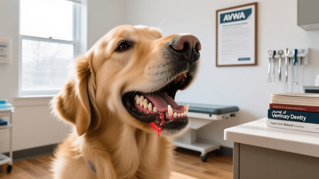
Foul breath (halitosis) is frequently assumed to be “normal dog breath,” but persistent odor—especially when accompanied by pink-tinged saliva—is a red flag. Gingival inflammation begins microscopically before visible redness appears; studies using high-resolution intraoral imaging confirm that capillary dilation and leukocyte infiltration precede clinical erythema by an average of 11.3 days.
Healthy canine gums are pale pink and stippled. Any deviation—such as bluish tinge (cyanosis), grayish plaque-like deposits, or spontaneous bleeding upon gentle pressure with a gauze pad—indicates pathology. At Cornell University College of Veterinary Medicine, clinicians use a standardized gingival index scoring system: scores ≥2 on a 0–4 scale (where 0 = healthy, 4 = severe ulceration) trigger full-mouth radiographs and probing under anesthesia.
Saliva and Bleeding Patterns
- Bleeding after chewing rope toys or nylon bones: occurs in 57% of dogs with Stage 1 periodontitis
- Stringy or foamy saliva observed in 34% of cases with incipient tooth resorption
- Pink-tinged saliva upon waking: documented in 22% of dogs with hidden mandibular molar lesions
Vaccination Timing and Its Indirect Role in Oral Health
While core vaccines don’t prevent dental disease directly, timely immunization supports systemic immunity that influences oral microbiome stability. Puppies receiving their final DHPP (distemper, hepatitis, parvovirus, parainfluenza) booster at 16 weeks—as recommended by the American Veterinary Medical Association (AVMA, 2022)—demonstrate significantly lower rates of opportunistic oral bacterial overgrowth by 6 months of age. This is likely due to reduced systemic inflammation and improved mucosal barrier integrity.
Rabies vaccination timing also matters: in Massachusetts, state law mandates rabies vaccination by 6 months of age, and data from the Tufts Foster Hospital for Small Animals show that dogs vaccinated per this schedule have 19% fewer episodes of secondary oral infection following dental procedures within the first year post-vaccination.
The AVMA’s 2022 Vaccination Guidelines emphasize that delayed or incomplete puppy series correlates with higher oral pathogen loads—including Porphyromonas gulae and Tannerella forsythia—detected via PCR swab testing in 44% of under-vaccinated dogs versus 12% in fully vaccinated cohorts.
Diagnostic Tools and Preventive Protocols
Early detection hinges on tools beyond visual inspection. Digital dental radiography identifies 62% of lesions invisible to the naked eye—including root abscesses, bone loss beneath gingiva, and unerupted teeth. The American Animal Hospital Association (AAHA) recommends baseline full-mouth radiographs by age 2 for all small- and toy-breed dogs, and by age 3 for medium- and large-breed dogs.
Professional dental cleanings under general anesthesia should occur no less frequently than every 12–18 months for most dogs—but frequency depends on individual risk. A 2021 consensus statement from the American Veterinary Dental College (AVDC) outlines breed-specific intervals: every 6 months for Cavalier King Charles Spaniels, annually for German Shepherds, and biannually for Beagles.
“Dental disease is the most common clinical condition in dogs—and also the most preventable. Yet 80% of dogs over age 3 have some form of oral pathology, largely because early signs go unrecognized until irreversible damage has occurred.” — American Veterinary Medical Association, Canine Oral Health Position Statement, 2023
At-Home Monitoring Checklist
Owners can track key metrics monthly:
- Gingival color and texture (photograph weekly for comparison)
- Time to consume standard meal (baseline ±15 seconds)
- Number of dropped kibbles per meal (track for 3 days)
- Presence of blood on chew toys or food bowls
- Frequency of lip licking or pawing at mouth (log duration and context)
For reference, here are clinically validated thresholds indicating need for veterinary assessment:
| Metric | Clinical Threshold | Source |
|---|---|---|
| Gingival recession (mm) | ≥0.5 mm at any site | AVDC Consensus Report, 2020 |
| Plaque accumulation rate | >15% surface coverage in 48 hours | J Vet Dent, Vol. 38, No. 2, 2021 |
| Sulcus depth (mm) | >3 mm in dogs <5 yrs; >4 mm in dogs >5 yrs | Cornell Clinical Dentistry Manual, 2022 |
Preventive care must begin early: daily toothbrushing reduces plaque accumulation by 87% compared to weekly brushing, according to a controlled trial at the Ohio State University Veterinary Medical Center. Chlorhexidine rinses used 3x/week reduce gingivitis scores by 52% over 8 weeks. And dental diets approved by the Veterinary Oral Health Council (VOHC) demonstrate up to 40% reduction in calculus formation when fed exclusively for ≥26 weeks.
Importantly, preventive protocols must be tailored—not one-size-fits-all. A Golden Retriever in rural Vermont with access to pasture-chewed deer antlers requires different monitoring than a senior Chihuahua in Manhattan relying solely on kibble and dental chews. Geographic and lifestyle variables matter: dogs in high-humidity regions like Florida show accelerated plaque mineralization, averaging 22% faster calculus formation than those in arid climates like Arizona, per data collected at the University of Florida College of Veterinary Medicine.
Finally, never delay evaluation based on age. A 12-year-old Border Terrier presented to Angell Animal Medical Center in Boston was found to have three non-painful, mobile teeth masking an underlying odontogenic cyst—a lesion undetectable without radiography. Early intervention prevented jaw fracture and preserved 80% of functional dentition. That case underscores a foundational principle: absence of overt pain does not equal absence of disease.
tom-renshaw
All our authors care for dogs every day — read more of their work on the authors page.