Pictures of Dog Tumors and Cysts
Learn to identify common dog tumors and cysts with visual descriptions, symptoms, diagnosis costs, treatment options, and when to see your vet.
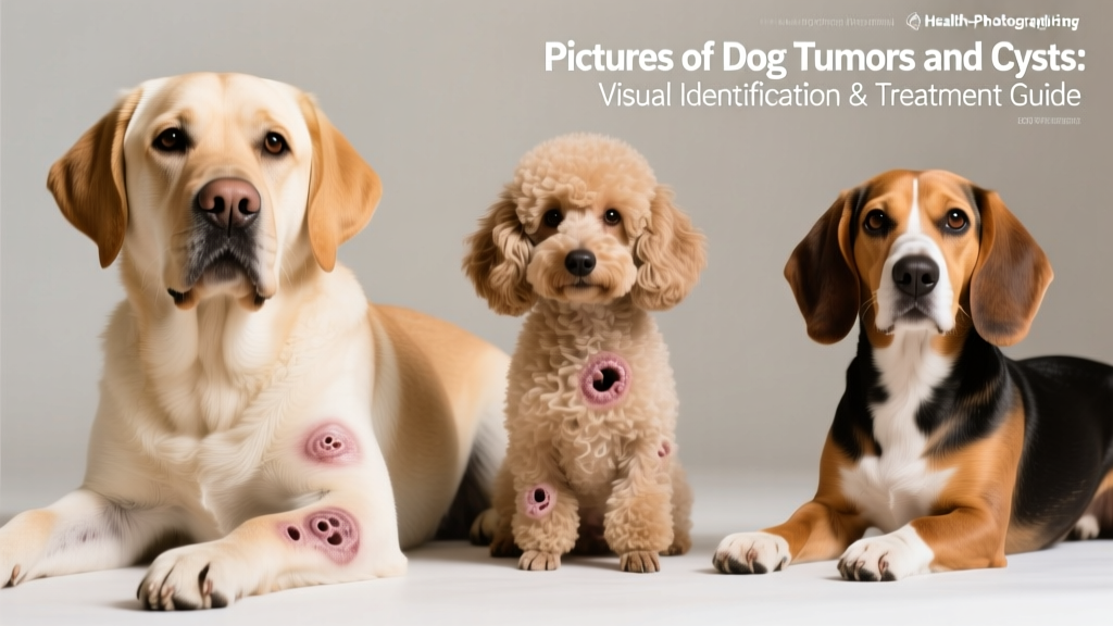
Finding a lump on your dog can trigger instant panic. While photographs and pictures of dog tumors and cysts can help you understand what you might be dealing with, no image replaces a veterinary diagnosis. Skin masses in dogs range from harmless sebaceous cysts to aggressive mast cell tumors, and many look nearly identical to the naked eye.
A tumor (neoplasm) is an abnormal growth of cells that can be benign (non-cancerous) or malignant (cancerous). A cyst is a fluid-filled or semi-solid sac beneath the skin, almost always benign. According to the American Kennel Club, approximately 30% of dogs over age 10 will develop at least one tumor during their lifetime, and skin tumors are the most commonly diagnosed neoplasms in dogs. Cysts are even more prevalent, with sebaceous cysts appearing in dogs of all ages but especially in middle-aged and senior animals.
Common Types of Dog Tumors and Cysts: Visual Descriptions
Since we cannot embed clinical photographs here, the following descriptions detail the visual characteristics that distinguish each type of mass. Use these as a reference when examining your dog, but remember that definitive diagnosis requires veterinary testing.
Sebaceous Cysts
These appear as smooth, round, dome-shaped bumps under the skin, typically 0.5 to 3 cm in diameter. They feel firm but slightly movable when gently pressed. The overlying skin is usually normal-colored or slightly bluish. Sometimes a small dark pore (punctum) is visible on top. If ruptured, they release a thick, white-to-yellow, toothpaste-like material (keratin and sebum). Most commonly found on the back, neck, and legs. Breeds prone to sebaceous cysts include Cocker Spaniels, Poodles, and German Shepherds.
Lipomas (Fatty Tumors)
Lipomas present as soft, squishy, freely movable lumps beneath the skin. They are not attached to deeper tissue and feel like a water balloon or soft dough. Size ranges from pea-sized to grapefruit-sized in extreme cases. The overlying skin looks completely normal. They are most common on the chest, abdomen, and upper legs. Overweight and older dogs are particularly susceptible. Infiltrative lipomas, a less common variant, feel firmer and are attached to underlying muscle.
Mast Cell Tumors
These are the great imitators of canine dermatology. A mast cell tumor can look like a raised pink bump, a hairless ulcerated patch, a soft lipoma-like mass, or an angry red welt. They may appear suddenly, change size daily (due to histamine release causing local swelling), and sometimes cause the surrounding skin to become red and inflamed — a phenomenon called Darier's sign. Found anywhere on the body but common on the trunk, limbs, and genital area. Boxers, Boston Terriers, Labrador Retrievers, and Pugs are predisposed.
Histiocytomas
These appear as small (usually under 2.5 cm), bright pink-to-red, hairless, button-like growths. They have a smooth or slightly ulcerated surface and tend to appear rapidly over one to two weeks. Most common in dogs under 3 years old, typically on the head, ears, and front legs. Most resolve spontaneously within two to three months, but veterinary confirmation is essential because they can resemble mast cell tumors.
Melanomas
Cutaneous melanomas appear as darkly pigmented (brown-to-black) raised masses, often with an irregular surface. They can be 1-5 cm and may be firm. Some are amelanotic (pink/flesh-colored), making them harder to identify visually. Benign melanomas are more common on haired skin; malignant melanomas tend to appear around the mouth, nail beds, and mucous membranes.
Papillomas (Warts)
These look like small, rough, cauliflower-like growths. They are usually pink or gray and appear in clusters around the mouth, eyes, or abdomen. Most common in puppies and immunocompromised senior dogs. Caused by canine papillomavirus, they typically resolve on their own within one to five months.
Symptom Checklist: Early Warning Signs vs. Advanced Indicators
Organizing symptoms by severity helps you determine urgency. Not every lump produces all of these signs, and some aggressive tumors show few external symptoms initially.
| Category | Early Warning Signs (Monitor Closely) | Advanced Symptoms (Vet Visit Needed) |
|---|---|---|
| Mass Appearance | Small, stable lump under 1 cm; smooth surface; normal skin color | Rapid growth (doubles in days/weeks); ulceration; bleeding; irregular shape; dark or mottled coloration |
| Texture & Mobility | Soft, freely movable beneath skin | Hard, fixed to underlying tissue; feels anchored or immovable |
| Behavioral Changes | Dog occasionally licks or scratches at area | Persistent licking/chewing; pain when touched; reluctance to be handled; decreased activity |
| Systemic Signs | None typically present at early stage | Weight loss; appetite decrease; vomiting; diarrhea; lethargy; enlarged lymph nodes |
| Discharge | Occasional clear fluid from cyst | Pus, blood, foul-smelling discharge; non-healing open sore |
- A lump that has grown noticeably within 48 hours
- Any mass that is bleeding, oozing pus, or has an open wound
- A firm, immovable lump attached to deeper tissue
- Multiple new lumps appearing simultaneously
- Lump accompanied by lethargy, vomiting, loss of appetite, or collapse
- Any mass near the eye, mouth, or genitals
- Swollen lymph nodes (lumps under jaw, behind knees, or in armpits)
Risk Factors: Which Dogs Are Most Vulnerable?
Key Risk Factors for Dog Tumors and Cysts
Age: Dogs over 7 years have significantly higher tumor risk. Cysts can occur at any age but peak in middle-aged dogs (5-10 years).
Breed Predisposition:
- Mast cell tumors: Boxers, Boston Terriers, Labrador Retrievers, Pugs, Golden Retrievers, Weimaraners
- Lipomas: Labrador Retrievers, Doberman Pinschers, Miniature Schnauzers, Cocker Spaniels, mixed breeds
- Sebaceous cysts: Cocker Spaniels, Poodles, German Shepherds, Huskies
- Histiocytomas: Scottish Terriers, Bulldogs, Boxers, Greyhounds (especially under age 3)
- Melanomas: Scottish Terriers, Airedale Terriers, Boston Terriers, Cocker Spaniels, Schnauzers
- Squamous cell carcinoma: Keeshonds, Standard Poodles, Norwegian Elkhounds, dogs with light-colored/short coats
Weight: Overweight dogs have higher lipoma incidence and increased overall cancer risk. A body condition score (BCS) above 6/9 correlates with higher tumor prevalence.
Genetics: Dogs with close relatives that developed tumors have elevated risk. Inbreeding and limited genetic diversity contribute.
Environment: Prolonged UV exposure increases squamous cell carcinoma and hemangiosarcoma risk, especially in white-coated and hairless areas. Chemical exposure (herbicides, pesticides) may elevate lymphoma and bladder cancer risk.
Sex & Reproductive Status: Intact females have higher mammary tumor risk (26% lifetime risk vs. 0.5% if spayed before first heat). Male dogs have higher perianal tumor rates.
How Vets Diagnose Dog Tumors and Cysts
Diagnosis follows a structured process. Your vet will not guess based on appearance alone — even experienced oncologists cannot reliably identify tumor types by sight. Here is what to expect:
- Physical Examination ($50-$100): The vet palpates the mass, checking size, shape, mobility, temperature, and attachment to underlying tissue. They examine regional lymph nodes for enlargement. A body condition score and general health assessment are recorded.
- Fine Needle Aspirate (FNA) / Cytology ($75-$200): A thin needle extracts cells from the mass, which are smeared onto a slide and examined under a microscope. This is the first-line diagnostic for most skin masses. Results may be available same-day if your vet has an in-house pathologist, or within 1-3 business days if sent to an external lab. FNA provides a definitive diagnosis in approximately 70-80% of cases. For cysts, the aspirate typically yields keratin debris or clear fluid, confirming the diagnosis immediately.
- Punch or Incisional Biopsy ($200-$500): If FNA results are inconclusive or suggest malignancy, a tissue biopsy is taken under local or general anesthesia. A pathologist examines the tissue architecture, providing a definitive diagnosis and tumor grade. Turnaround: 5-10 business days. This is the gold standard for mast cell tumor grading (Grade I, II, or III), which directly determines treatment and prognosis.
- Blood Work and Urinalysis ($120-$300): A complete blood count (CBC), chemistry panel, and urinalysis assess organ function, check for systemic disease, and establish a baseline before any treatment. Elevated liver enzymes or abnormal white blood cell counts may indicate metastasis or paraneoplastic syndromes.
- Imaging — X-rays, Ultrasound, or CT ($200-$1,500): Thoracic radiographs (chest X-rays, $150-$350) check for lung metastasis. Abdominal ultrasound ($300-$600) evaluates internal organs and lymph nodes. CT scans ($800-$1,500) provide detailed 3D imaging for surgical planning, especially for masses on the limbs or near critical structures.
- Staging (for malignant tumors): Staging determines how far cancer has spread. It typically combines FNA/biopsy of the primary mass, lymph node aspirates, chest X-rays, and abdominal ultrasound. Total staging cost: $600-$2,000 depending on complexity.
Treatment Options: What Works for Each Type of Mass
| Mass Type | Primary Treatment | Cost Range | Success Rate / Outcome |
|---|---|---|---|
| Sebaceous Cyst | Surgical excision if problematic; monitor if small and stable. Do not squeeze or lance at home. | $200-$600 (surgical removal) | Curative with complete excision. Recurrence possible if capsule not fully removed. |
| Lipoma | Usually no treatment needed. Surgical removal if mass restricts movement, grows rapidly, or causes discomfort. | $300-$800 (surgical removal); $1,000-$2,500 (infiltrative lipoma) | Excellent. Simple lipomas rarely recur after removal. Infiltrative lipomas have higher recurrence (30-50%). |
| Mast Cell Tumor | Wide surgical excision (2-3 cm margins). Chemotherapy (vinblastine/prednisone) for Grade II-III or incomplete margins. Radiation for non-resectable tumors. | $1,000-$3,000 (surgery); $3,000-$8,000+ (chemotherapy protocol); $4,000-$8,000 (radiation) | Grade I: >90% cure with surgery. Grade II: 50-70% long-term survival with surgery + chemo. Grade III: median survival 4-6 months without treatment, up to 12-18 months with aggressive therapy. |
| Histiocytoma | Usually self-resolving within 2-3 months. Surgical removal if persistent, ulcerated, or diagnosis uncertain. | $0 (watchful waiting); $200-$600 (if removal needed) | Excellent. Self-limiting in most cases. Rarely recurs. |
| Melanoma (Cutaneous) | Surgical excision. Melanoma vaccine (Oncept) for malignant variants. Radiation or chemotherapy for oral/digital melanomas. | $800-$2,500 (surgery); $1,500-$3,000 (vaccine series); $4,000-$10,000+ (comprehensive treatment) | Benign cutaneous: curative with surgery. Malignant oral/digital: median survival 5-8 months without treatment, 12-18 months with surgery + vaccine. |
| Papilloma (Warts) | Self-resolving in immunocompetent dogs. Surgical removal or cryotherapy if persistent or obstructive. Azithromycin sometimes prescribed. | $0 (self-resolving); $150-$400 (surgical/cryo) | Excellent. Most resolve within 1-5 months without intervention. |
Supportive and Adjunctive Care
Beyond primary treatment, supportive care improves quality of life and outcomes:
- Pain management: NSAIDs (carprofen, meloxicam) at $30-$80/month; gabapentin for neuropathic pain at $15-$40/month
- Nutritional support: Prescription oncology diets (Hill's Prescription Diet n/d, Royal Canin Recovery) at $50-$90/bag; omega-3 fatty acid supplementation (fish oil, 1,000-3,000 mg EPA/DHA daily depending on dog size)
- Immune support: Turkey tail mushroom extract (I'm-Yunity, polysaccharopeptide-based) showed promise in a University of Pennsylvania study for hemangiosarcoma — discuss with your veterinary oncologist
- Wound care: Post-surgical sites require daily monitoring, Elizabethan collar use (cone), and possible bandage changes for 10-14 days
Prognosis: What to Expect by Mass Type and Stage
| Mass Type | Best-Case Scenario | Worst-Case Scenario | Key Prognostic Factor |
|---|---|---|---|
| Sebaceous Cyst | Normal lifespan; may never require treatment | Infection or rupture causing local inflammation; rarely life-threatening | Location and whether the cyst ruptures |
| Lipoma | Normal lifespan; many dogs live with untreated lipomas for years | Infiltrative lipoma causing mobility issues; extremely rare malignant transformation (liposarcoma) | Whether lipoma is simple or infiltrative |
| Mast Cell Tumor | Grade I: full cure, normal lifespan | Grade III with metastasis: median survival 2-4 months without treatment | Tumor grade (I, II, or III), mitotic index, KIT mutation status, surgical margins |
| Histiocytoma | Self-resolving; no long-term impact | Rarely: systemic histiocytosis in Bernese Mountain Dogs (life-threatening) | Correct diagnosis (must rule out mast cell tumor) |
| Melanoma | Benign cutaneous: curative with surgery | Malignant oral: 6-12 month survival even with treatment | Location (skin vs. oral vs. nail bed), mitotic index |
| Papilloma | Self-resolving within months | Persistent in immunocompromised dogs; rarely undergoes malignant transformation | Dog's immune status |
According to the Merck Veterinary Manual, the single most important prognostic factor for mast cell tumors is histologic grade. Dogs with Grade I tumors treated with complete surgical excision have a greater than 90% long-term survival rate, while Grade III tumors carry a significantly guarded prognosis even with aggressive treatment.
Home Care and Daily Management
Monitoring Existing Masses
If your vet has diagnosed a benign mass and recommended watchful waiting, follow this protocol:
- Weekly measurements: Use a ruler or calipers to measure the mass in two dimensions (length and width in millimeters). Record in a notebook or phone app with the date.
- Monthly photographs: Take a photo with a coin or ruler for scale, under consistent lighting. Compare month-to-month for color, shape, or surface changes.
- Palpation checks: Gently feel the mass weekly. Note changes in firmness, mobility, temperature (warmth may indicate inflammation or infection), and whether your dog reacts to touch.
- Track behavior: Note any new licking, scratching, or chewing at the site. An Elizabethan collar ($10-$25) can prevent self-trauma while you arrange a vet visit.
Post-Surgical Care
After mass removal, expect a 10-14 day recovery period:
- Keep the incision clean and dry — no bathing for 10-14 days
- Elizabethan collar at all times to prevent licking (suture infection is the most common post-op complication)
- Restrict activity: leash walks only, no running, jumping, or rough play
- Check the incision twice daily for redness, swelling, discharge, or dehiscence (opening)
- Administer prescribed pain medication and antibiotics on schedule
- Return for suture removal at 10-14 days (if non-absorbable sutures were used)
Lifestyle Adjustments
For dogs with cancer diagnoses, quality of life is paramount:
- Nutrition: Feed a calorie-dense, high-protein diet. Cancer increases metabolic demands. Discuss specific formulations with your vet.
- Exercise: Maintain gentle daily walks (15-30 minutes) to preserve muscle mass and mental wellbeing. Adjust intensity to your dog's energy level.
- Comfort: Orthopedic beds ($40-$150) reduce pressure on surgical sites and improve rest quality. Ramps for furniture and car access prevent strain.
- Quality of life tracking: Use the HHHHHMM scale (Hurt, Hunger, Hydration, Hygiene, Happiness, Mobility, More good days than bad) — a validated tool for assessing end-of-life quality. Score each category 1-10 daily; a total below 35 warrants discussion with your vet.
Complete Cost Breakdown: Diagnosis Through Treatment
| Service | Low Estimate | High Estimate | Notes |
|---|---|---|---|
| Initial exam + consultation | $50 | $150 | Higher at specialty/emergency clinics |
| Fine needle aspirate (FNA) | $75 | $200 | In-house cytology is cheaper |
| Biopsy (punch or incisional) | $200 | $500 | Includes pathology lab fee |
| Blood work + urinalysis | $120 | $300 | CBC, chemistry panel, UA |
| Chest X-rays (3 views) | $150 | $350 | For metastasis screening |
| Abdominal ultrasound | $300 | $600 | Specialist referral may cost more |
| Benign mass removal (cyst/lipoma) | $200 | $800 | Varies by mass size and location |
| Malignant tumor surgery | $1,000 | $4,000 | Wide margins, complex closures cost more |
| Chemotherapy (full protocol) | $3,000 | $10,000+ | Depends on drug protocol and number of cycles |
| Radiation therapy | $4,000 | $8,000 | Typically 10-20 fractions |
| Total: Benign mass workup + removal | $445 | $1,950 | |
| Total: Malignant tumor full treatment | $4,895 | $25,000+ | Worst-case with surgery + chemo + radiation |
- Pet insurance: If enrolled before diagnosis, most policies cover 70-90% of cancer treatment costs after deductible. Premiums average $30-$70/month for comprehensive coverage in 2026.
- CareCredit: Healthcare credit line offering 0% promotional financing for 6-24 months on veterinary bills over $200.
- Charitable assistance: Organizations like the Brown Dog Foundation, RedRover Relief, and the Magic Bullet Fund offer grants for cancer treatment.
- Teaching hospitals: University veterinary teaching hospitals often offer 20-40% lower specialist fees while providing cutting-edge care.
Prevention Strategies
While not all tumors are preventable, these evidence-based strategies reduce risk:
- Early spay/neuter: Spaying females before their first heat cycle reduces mammary tumor risk from 26% to less than 0.5%. Neutering males eliminates testicular cancer and reduces perianal adenoma risk by over 90%.
- Weight management: Maintain a body condition score of 4-5/9. Obesity is a confirmed cancer risk factor in dogs, linked to increased incidence of lipomas, mammary tumors, and transitional cell carcinoma. According to the AVMA, approximately 56% of dogs in the US are overweight or obese.
- Sun protection: Apply dog-safe sunscreen (zinc oxide-free, SPF 30+) to lightly pigmented areas — nose, ear tips, belly — for dogs that spend extended time outdoors. Dog-specific sunscreens like Epi-Pet or My Dog Nose It cost $10-$20 per tube.
- Limit chemical exposure: Wash your dog's paws after walking on treated lawns. Store herbicides and pesticides securely. A 2025 study in the Journal of Veterinary Internal Medicine found dogs exposed to lawn herbicides had elevated bladder cancer risk.
- Regular wellness exams: Biannual veterinary check-ups catch masses early when treatment is most effective and least expensive. Ask your vet to perform a full-body palpation at every visit.
- Home body checks: Once a month, run your hands over your dog's entire body — head to tail, including between toes, inside ears, under the tail, and along the belly. Familiarity with your dog's normal bumps lets you spot changes quickly.
Frequently Asked Questions
Can I tell if a dog's lump is cancerous by looking at pictures?
No. While pictures of dog tumors and cysts can help you understand general categories, many benign and malignant masses look identical visually. Mast cell tumors, for example, can mimic lipomas. Only cytology (FNA) or biopsy can provide a definitive diagnosis. If you find a lump, schedule a veterinary visit rather than relying on image comparison.
How fast do dog tumors grow?
Growth rates vary enormously. Histiocytomas can appear overnight and resolve in weeks. Low-grade mast cell tumors may grow slowly over months. High-grade mast cell tumors and sarcomas can double in size within days. Any mass that changes noticeably in 1-2 weeks warrants immediate veterinary evaluation.
Should I squeeze or drain my dog's cyst at home?
Absolutely not. Squeezing a cyst can cause rupture beneath the skin, triggering a severe inflammatory response, infection, and abscess formation. This makes eventual surgical removal more difficult and expensive. Leave drainage to your veterinarian, who will use sterile technique and determine whether the cyst actually needs intervention.
What is the life expectancy of a dog with a tumor?
This depends entirely on tumor type, grade, stage, and treatment. Dogs with benign tumors (lipomas, sebaceous cysts, histiocytomas) have a normal life expectancy. Dogs with low-grade mast cell tumors treated surgically often live normal lifespans. High-grade malignancies with metastasis may carry survival times of 2-6 months without treatment, extending to 12-18 months with aggressive therapy. Your veterinary oncologist can provide specific prognosis based on your dog's individual diagnosis.
Are certain breeds more prone to tumors and cysts?
Yes. Boxers, Boston Terriers, and Pugs have notably high mast cell tumor rates. Labrador Retrievers are predisposed to both lipomas and mast cell tumors. Cocker Spaniels and Poodles frequently develop sebaceous cysts. Bernese Mountain Dogs, Golden Retrievers, and Rottweilers have elevated overall cancer rates. Mixed-breed dogs generally have lower cancer risk due to greater genetic diversity (hybrid vigor), though they are not immune.
How much does it cost to have a lump checked?
A basic lump workup — exam plus FNA — typically costs $125-$350 at a general practice. If biopsy, blood work, and imaging are needed, total diagnostic costs range from $500-$2,000. Early diagnosis is significantly cheaper than treating advanced disease.
When should I see the vet about a lump on my dog?
Schedule a non-urgent appointment for any new lump you discover, ideally within one to two weeks. Seek urgent care (same-day or emergency) if the mass is rapidly enlarging, bleeding, ulcerated, causing pain, or accompanied by systemic symptoms like lethargy, appetite loss, vomiting, or swollen lymph nodes.
The Bottom Line
Finding a lump on your dog is frightening, but the majority of skin masses in dogs are benign or highly treatable when caught early. The single most important action you can take is having any new mass evaluated by your veterinarian through FNA cytology — a quick, minimally invasive, and relatively affordable test that provides critical information. Do not rely on pictures of dog tumors and cysts for diagnosis. Instead, use this guide as a framework for understanding what your vet finds and making informed decisions about your dog's care.
This article is for educational purposes and does not replace professional veterinary advice. Always consult your veterinarian for diagnosis and treatment of any health condition in your dog.
Priya Sutaria
All our authors care for dogs every day — read more of their work on the authors page.