Dog Skin Cancer: Signs, Types, Treatment & Prognosis Guide
Comprehensive guide to dog skin cancer covering types, symptoms by severity, diagnosis costs, treatment options, survival rates, and breed risks.
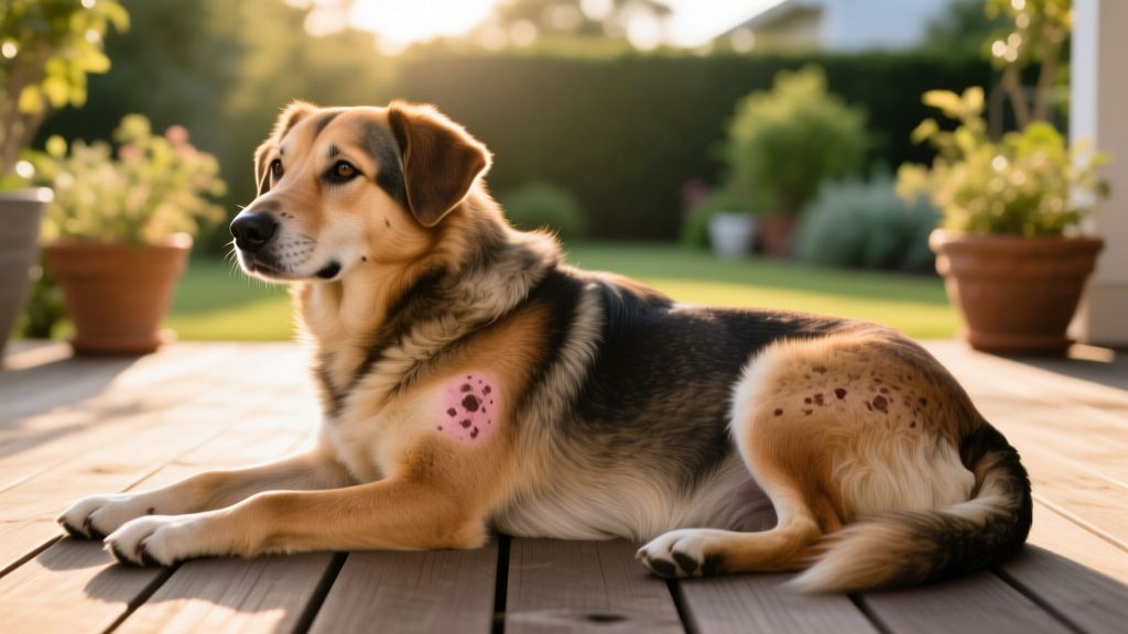
Skin cancer is the most commonly diagnosed tumor type in dogs, accounting for roughly 30% of all canine cancers according to the Merck Veterinary Manual. It refers to any uncontrolled growth of abnormal cells originating in the skin layers — the epidermis, dermis, or subcutaneous tissue. These growths can be benign (non-spreading) or malignant (invasive and potentially metastatic). UV radiation, genetics, chronic inflammation, and certain viral infections all contribute to development. While any dog can develop skin cancer, incidence rises sharply after age seven, and some breeds carry significantly elevated genetic risk.
Early detection dramatically improves outcomes. Many skin cancers caught at Stage I carry cure rates above 80% with surgical removal, while advanced metastatic disease has a far graver prognosis. This guide walks you through the types, warning signs, diagnostic process, treatment pathways, and real costs you should expect.
What Is Skin Cancer in Dogs? Understanding the Major Types
Canine skin cancer is not a single disease — it's an umbrella term covering dozens of distinct tumor types, each with different behaviors, treatments, and outcomes. The three most clinically significant malignant types are:
Mast Cell Tumors (MCTs): The most common malignant skin tumor in dogs, representing approximately 16–21% of all skin tumors. MCTs originate from mast cells in the dermis and can range from low-grade (slow-growing, rarely metastasize) to high-grade (aggressive, high metastatic potential). They often appear as raised, firm lumps but can mimic benign growths, earning them the nickname "the great imitator" among veterinary oncologists.
Melanomas: Arising from pigment-producing melanocytes, cutaneous melanomas in dogs are actually more often benign than malignant — the opposite of human melanoma. However, malignant melanomas that do develop on haired skin can be aggressive. Mucosal melanomas (mouth, nail bed) are far more dangerous and carry high metastatic rates.
Squamous Cell Carcinomas (SCC): These tumors develop from the squamous epithelial cells and are strongly linked to UV exposure in lightly pigmented, sparsely haired areas like the belly, nose, and ear tips. They tend to be locally invasive but metastasize relatively slowly.
Other notable types include fibrosarcomas, hemangiosarcomas of the skin, histiocytomas (typically benign and self-resolving in young dogs), and sebaceous gland adenomas (almost always benign).
Symptoms Checklist: Early Warning Signs vs. Advanced Disease
Skin cancer symptoms vary enormously depending on tumor type, location, and stage. Use this checklist to categorize what you're observing and determine urgency.
| Severity Level | Symptoms | Action Required |
|---|---|---|
| Early (Monitor Closely) |
• New lump or bump persisting beyond 2–3 weeks • Small raised area, firm or rubbery to touch • Pigmented spot that has changed shape or color • Hairless patch over a subcutaneous nodule • Single lesion less than 1 cm in diameter |
Schedule routine vet appointment within 1–2 weeks |
| Moderate (Prompt Evaluation) |
• Rapidly growing mass (noticeable change in days to weeks) • Ulcerated or bleeding skin lesion that won't heal • Lump that is fixed to underlying tissue (doesn't move freely) • Multiple new masses appearing simultaneously • Redness, warmth, or swelling around a lesion • Mass larger than 2 cm • Dog licking, scratching, or biting at a specific spot persistently |
Vet appointment within 48–72 hours |
| Advanced (Urgent/Immediate) |
• Large, fungating (cauliflower-like) or necrotic wound • Enlarged regional lymph nodes (especially submandibular or popliteal) • Unexplained weight loss or muscle wasting • Lethargy, decreased appetite, vomiting • Difficulty breathing or persistent cough • Bleeding that doesn't stop with pressure • Sudden collapse or inability to stand |
Emergency vet visit immediately |
The "1-2-3 Rule" for lump evaluation: Many veterinary dermatologists recommend this framework — if a lump is 1 cm or larger, has been present for 2 weeks or more, or is 3 months old regardless of size, it warrants professional evaluation with fine needle aspiration (FNA) at minimum.
Risk Factors: Which Dogs Are Most Vulnerable?
Breed Predisposition
Certain breeds have well-documented elevated risk for specific skin cancers:
- Boxers, Boston Terriers, Bulldogs, Pugs: High risk for mast cell tumors — Boxers carry a documented genetic mutation (c-KIT) linked to MCT development
- Scottish Terriers, Schnauzers, Doberman Pinschers: Elevated melanoma risk
- White or lightly pigmented breeds (Dalmatians, Bull Terriers, Beagles, Whippets): Higher squamous cell carcinoma risk due to UV susceptibility
- Golden Retrievers, Labrador Retrievers: Above-average rates of multiple skin tumor types
- Shar-Peis: Predisposed to cutaneous histiocytomas and mast cell tumors
Non-Breed Risk Factors
- Age: Median age for MCT diagnosis is 8–9 years; SCC median is 9–10 years. Risk increases substantially after age 7.
- Sun exposure: Dogs with short, light-colored coats and minimal abdominal pigmentation face the highest UV-related SCC risk.
- Chronic skin inflammation: Long-standing dermatitis, hot spots, or lick granulomas may create a pro-carcinogenic microenvironment.
- Immunosuppression: Dogs on long-term corticosteroids or with immune-mediated conditions may have reduced tumor surveillance.
- Weight: Obesity is associated with increased cancer risk across multiple tumor types in dogs, though the mechanism is not fully understood.
How Is Dog Skin Cancer Diagnosed? Tests, Timelines & Costs
The diagnostic pathway typically follows a staged approach, escalating in complexity based on initial findings.
Diagnostic Process Flow
- Physical Examination & History ($50–$95 office visit fee)
Your vet will palpate the mass, assess mobility, measure dimensions, check regional lymph nodes, and perform a full-body skin survey to identify any additional lesions. - Fine Needle Aspiration / Cytology ($75–$200)
A thin needle extracts cells from the mass, which are smeared onto slides and examined under a microscope — often in-clinic or sent to a reference lab. This is the single most important first test. It can identify mast cell tumors, melanomas, lipomas, and many other masses with 70–90% accuracy depending on tumor type. Results typically available within 24–72 hours. - Surgical Biopsy & Histopathology ($200–$500)
If FNA results are inconclusive or if tumor grading is needed (critical for MCTs), a tissue sample or the entire mass is removed and sent to a veterinary pathologist. Histopathology provides the definitive diagnosis, tumor grade (I, II, or III for MCTs), and surgical margin assessment. Turnaround: 5–10 business days. - Staging Workup — if malignancy confirmed ($400–$1,200+)
- Complete blood count (CBC) and biochemistry panel: $150–$300
- Thoracic radiographs (3 views): $150–$350
- Abdominal ultrasound: $300–$600
- Regional lymph node aspiration: $75–$150
- Advanced imaging (CT or MRI) if available: $1,000–$2,500
- Specialized Testing (as indicated)
- Ki67 proliferation index for MCTs: $150–$300 — helps predict biological behavior of Grade II tumors
- c-KIT mutation testing: $200–$400 — relevant for targeted therapy decisions
- Immunohistochemistry panels: $200–$500 — used when tumor origin is unclear
Total initial diagnostic cost range: $200–$400 for a straightforward FNA-confirmed benign mass, or $800–$2,500+ for a full staging workup on a confirmed malignancy.
Treatment Options: Surgery, Medical Therapy & Supportive Care
Treatment selection depends on tumor type, grade, stage, location, and your dog's overall health. Here is a comparison of the primary modalities:
| Treatment | Best For | Cost Range | Success / Cure Rate | Side Effects |
|---|---|---|---|---|
| Surgical Excision | Localized, non-metastatic tumors; first-line for most skin cancers | $500–$3,000 (varies by size, location, reconstruction needs) | Grade I MCTs: >90% cure with clean margins; Grade II: 70–90%; SCC: 80–95% if completely excised | Wound healing complications, infection, seroma formation; 10–14 day recovery |
| Radiation Therapy | Incomplete surgical margins, non-resectable tumors, or as adjuvant post-surgery | $3,000–$8,000 (full course, 10–20 fractions) | Adjuvant for Grade II MCTs: 85–95% local control at 1 year; SCC: 70–85% response | Skin erythema, moist desquamation, temporary hair loss in treatment field; requires general anesthesia per session |
| Chemotherapy | High-grade, metastatic, or systemic disease | $2,000–$6,000 (varies by protocol and duration) | Variable — extends survival but rarely curative alone for advanced disease; vinblastine + prednisone for MCT: median survival 7–13 months for Grade III | GI upset, bone marrow suppression (monitored via CBC), rare but possible severe neutropenia |
| Tyrosine Kinase Inhibitors (e.g., Palladia/toceranib) | Mast cell tumors with c-KIT mutations, non-resectable MCTs | $200–$600/month ongoing | Objective response rate ~37–47% for MCTs; disease stabilization in additional 30–40% | GI signs, hypertension, proteinuria, muscle cramping; requires regular bloodwork monitoring |
| Cryosurgery / Electrocautery | Small, superficial benign or low-grade lesions | $200–$800 per lesion | High for small superficial benign tumors; not appropriate for malignant or deep lesions | Local tissue necrosis, sloughing over 1–3 weeks; minimal systemic effects |
| Immunotherapy (Melanoma Vaccine — Oncept) | Stage II–III oral/digital melanoma post-surgery | $1,500–$3,000 (initial 4-dose series + boosters) | Extends median survival time significantly when combined with surgery; not curative alone | Injection site reactions, mild fever; generally well tolerated |
Supportive care runs parallel to active treatment: pain management (NSAIDs like carprofen at $30–$80/month, or gabapentin at $15–$50/month), anti-nausea medications (maropitant/Cerenia), appetite stimulants, and wound care supplies. Never administer human NSAIDs (ibuprofen, naproxen) to your dog — these are toxic and can cause fatal gastrointestinal ulceration and kidney failure.
Prognosis by Stage and Tumor Type
Prognosis varies enormously based on tumor type, histologic grade, and stage at diagnosis. The following table reflects median survival times reported in veterinary oncology literature:
| Tumor Type & Grade | Stage / Description | Median Survival Time (with treatment) | Key Prognostic Factors |
|---|---|---|---|
| Mast Cell Tumor — Grade I | Localized, completely excised | >4 years (essentially normal lifespan; considered cured) | Clean surgical margins; low Ki67 index |
| Mast Cell Tumor — Grade II | Localized, completely excised | 2–4+ years (varies widely by sub-grading) | Mitotic index, Ki67, c-KIT pattern; high-grade Grade II behaves like Grade III |
| Mast Cell Tumor — Grade III | High-grade, with or without metastasis | 4–7 months (surgery alone); 7–13 months (with chemotherapy) | Lymph node involvement, liver/spleen metastasis, response to chemo |
| Cutaneous Melanoma (benign) | Localized, haired skin | Normal lifespan (cured with excision) | Histopathology confirmation of benign nature |
| Malignant Melanoma (cutaneous) | Locally invasive, no metastasis | 12–24 months with wide surgical excision | Mitotic index, location, size at diagnosis |
| Squamous Cell Carcinoma | Localized, completely excised | >2–3 years (low metastatic rate; local recurrence is main concern) | Tumor size, completeness of excision, digital vs. cutaneous location |
| Hemangiosarcoma (cutaneous) | Dermal (sun-induced) | >2 years with excision (much better than visceral HSA) | Dermal vs. subcutaneous — subcutaneous has much worse prognosis (~6 months) |
Important context: These are population-level medians. Individual dogs may do significantly better or worse depending on overall health, treatment response, and tumor biology. Your veterinary oncologist will provide a personalized prognosis based on your dog's specific pathology results.
Home Care, Monitoring & Lifestyle Adjustments
Whether your dog is undergoing active treatment or being monitored for a low-risk lesion, consistent home care plays a critical role in outcomes.
Daily Monitoring Protocol
- Weekly full-body skin checks: Part the coat systematically — head to tail, including belly, armpits, between toes, and inside ear flaps. Use your fingertips to feel for subcutaneous lumps that aren't visible.
- Document changes: Photograph any known masses monthly with a ruler or coin for scale. Note changes in size, color, texture, ulceration, or bleeding.
- Track behavior: Monitor appetite, energy level, water intake, and bathroom habits. Sudden changes may indicate systemic disease progression.
Post-Surgical Care
- Prevent licking/chewing at incision sites with an Elizabethan collar or recovery suit — wound dehiscence is a leading post-surgical complication.
- Administer all prescribed pain medications and antibiotics on schedule.
- Restrict activity for 10–14 days; no running, jumping, or rough play.
- Check incision daily for redness, swelling, discharge, or separation.
- Follow up for suture removal at 10–14 days (if non-absorbable sutures were used).
Sun Protection for High-Risk Dogs
For dogs with SCC risk factors (white coats, thin hair, sun-loving behavior):
- Apply dog-specific sunscreen (zinc oxide-free, as zinc oxide is toxic if ingested) to exposed areas — noses, ear tips, belly. Products like Epi-Pet Sunscreen or Doggles dog-specific sunblock are formulated for canine use.
- Limit outdoor time between 10 AM and 4 PM during peak UV months.
- Consider UV-protective bodysuits (e.g., Sunsuit by Doggles) for extended outdoor exposure.
Nutrition & Immune Support
While no diet prevents cancer, maintaining optimal body condition (BCS 4–5 out of 9) and providing balanced nutrition supports immune function. Discuss omega-3 fatty acid supplementation (EPA/DHA at 50–100 mg/kg combined daily) with your vet, as some evidence suggests anti-inflammatory and potential anti-neoplastic benefits in dogs.
How Much Does Dog Skin Cancer Treatment Cost? Full Breakdown
Treatment costs vary significantly based on tumor type, stage, geographic location, and whether you pursue care at a general practice or specialty oncology center. Here is a realistic budget framework:
| Expense Category | Low Estimate | High Estimate | Notes |
|---|---|---|---|
| Initial diagnostics (FNA, bloodwork, imaging) | $200 | $2,500 | Higher end includes full staging with ultrasound and CT |
| Surgical removal | $500 | $5,000 | Complex reconstructions or specialist surgery increase cost |
| Radiation therapy (full course) | $3,000 | $8,000 | Requires specialty center; varies by number of fractions |
| Chemotherapy (full protocol) | $2,000 | $6,000 | Vinblastine-based protocols for MCTs; includes monitoring labs |
| Targeted therapy (Palladia) | $200/mo | $600/mo | Ongoing; dose-dependent on body weight |
| Ongoing monitoring (rechecks, labs) | $100–$200/visit | $400–$600/visit | Every 3–6 months post-treatment; more frequent during active therapy |
| TOTAL (localized, surgery-only scenario) | $1,000 | $4,000 | Most common scenario for early-stage tumors |
| TOTAL (multimodal — surgery + radiation + chemo) | $6,000 | $15,000+ | Advanced or high-grade disease requiring comprehensive approach |
Financial planning tips: Pet insurance policies purchased before diagnosis typically cover 70–90% of oncology costs (minus deductible). If your dog is uninsured, ask your vet about CareCredit or Scratchpay payment plans. Many veterinary schools also offer reduced-cost oncology consultations through their teaching hospitals.
Prevention Strategies: Reducing Your Dog's Risk
Not all cancers are preventable — genetics play a substantial role — but several evidence-informed strategies can reduce risk:
- Minimize UV exposure for susceptible breeds: Use canine sunscreen on unpigmented areas, provide shade, and avoid midday sun for white or thin-coated dogs.
- Maintain ideal body weight: Obesity-associated chronic inflammation may promote tumorigenesis. Keep your dog at BCS 4–5/9.
- Perform regular skin checks: Monthly full-body palpation catches tumors at smaller sizes when surgical cure is most achievable.
- Address chronic skin disease promptly: Persistent inflammation from allergies, infections, or lick granulomas should be managed with veterinary guidance rather than left untreated for years.
- Discuss spay/neuter timing with your vet: Some research suggests intact dogs may have different cancer risk profiles depending on breed and sex. The AKC provides breed-specific guidance on optimal timing.
- Consider breed-specific genetic testing: For breeds with known cancer predispositions (e.g., Boxers with c-KIT mutations), genetic screening through services like Embark or Wisdom Panel can inform monitoring intensity.
When to See the Vet: Red Flags Requiring Immediate Attention
🚨 Emergency and Urgent Care Triggers
Seek immediate veterinary care (emergency clinic if after hours) if your dog exhibits:
- A mass that is bleeding profusely and won't stop with 5 minutes of direct pressure
- Sudden collapse, weakness, or inability to stand
- Difficulty breathing, rapid breathing, or persistent coughing
- Acute vomiting or diarrhea, especially with blood
- Sudden severe lethargy or unresponsiveness
- Rapid abdominal distension (could indicate internal bleeding from metastatic disease)
Schedule a prompt appointment (within 48–72 hours) for:
- Any new lump that is rapidly growing
- A mass that has ulcerated, opened, or is oozing
- Enlarged lymph nodes (firm swellings under the jaw, in front of shoulders, behind knees)
- Unexplained weight loss of more than 5% body weight
- Persistent licking or scratching at one specific area
- A previously stable lump that suddenly changes in size, shape, or color
If you suspect your dog has ingested a toxic substance (including topical chemotherapy medications, human NSAIDs, or toxic plants), contact the ASPCA Animal Poison Control Center at (888) 426-4435 (24/7, consultation fee applies) or the Pet Poison Helpline at (855) 764-7661.
Frequently Asked Questions
What is skin cancer in dogs?
Dog skin cancer encompasses any malignant tumor arising from the skin's cellular layers. The most common types are mast cell tumors, melanomas, squamous cell carcinomas, and fibrosarcomas. These tumors develop when genetic mutations cause skin cells to divide uncontrollably, forming masses that may invade surrounding tissue or spread to distant organs.
What are the symptoms of skin cancer in dogs?
Symptoms range from a simple raised lump to ulcerated, bleeding wounds. Early signs include any new or changing mass, pigmented skin lesions, or firm subcutaneous nodules. Advanced signs include enlarged lymph nodes, weight loss, lethargy, and non-healing wounds. Because many benign growths mimic malignant ones, any persistent lump warrants veterinary evaluation.
How is skin cancer diagnosed in dogs?
Diagnosis begins with a physical exam and fine needle aspiration (FNA) cytology, which provides a preliminary diagnosis in most cases. Definitive diagnosis and tumor grading require surgical biopsy with histopathology. If malignancy is confirmed, staging (bloodwork, chest X-rays, abdominal ultrasound, lymph node evaluation) determines whether the cancer has spread.
Can dog skin cancer be cured?
Yes — many skin cancers are curable when caught early and completely excised. Low-grade mast cell tumors, benign melanomas, and localized squamous cell carcinomas often carry excellent long-term prognosis with surgery alone. High-grade or metastatic tumors are more challenging but can still be managed to extend quality of life significantly. The key variable is stage at diagnosis.
How much does skin cancer treatment cost for dogs?
Costs range from approximately $1,000–$4,000 for straightforward surgical removal of a localized tumor, to $6,000–$15,000+ for multimodal treatment (surgery, radiation, and chemotherapy) of advanced disease. Pet insurance, veterinary school hospitals, and payment plans can significantly reduce out-of-pocket expenses.
What is the life expectancy of a dog with skin cancer?
Life expectancy ranges from a normal full lifespan (for benign tumors or completely excised low-grade malignancies) to 4–7 months for high-grade metastatic mast cell tumors without treatment. With aggressive multimodal therapy, even high-grade tumors may see survival times of 7–13 months or longer. Your veterinary oncologist will provide the most accurate estimate based on your dog's specific pathology.
Which breeds are prone to skin cancer?
Boxers, Boston Terriers, Bulldogs, and Pugs have the highest mast cell tumor risk. Scottish Terriers and Schnauzers face elevated melanoma risk. White or lightly pigmented breeds (Dalmatians, Bull Terriers, Whippets) are predisposed to UV-related squamous cell carcinoma. Golden Retrievers and Labrador Retrievers show above-average rates across multiple skin tumor types.
Are lumps on dogs always cancer?
No. In fact, the majority of skin masses in dogs are benign — lipomas (fatty tumors), sebaceous adenomas, histiocytomas, and skin tags are extremely common. However, because malignant tumors can closely resemble benign ones visually and even on palpation, the only reliable way to distinguish them is through veterinary cytology (FNA) or biopsy. The AVMA recommends having all new or changing lumps evaluated.
Can I prevent my dog from getting skin cancer?
You cannot eliminate risk entirely — genetics play a major role — but you can reduce it. Minimize sun exposure for light-coated breeds, maintain a healthy weight, manage chronic skin conditions promptly, and perform monthly full-body skin checks to catch any tumors at their smallest and most treatable size.
Jonas Cole
All our authors care for dogs every day — read more of their work on the authors page.


