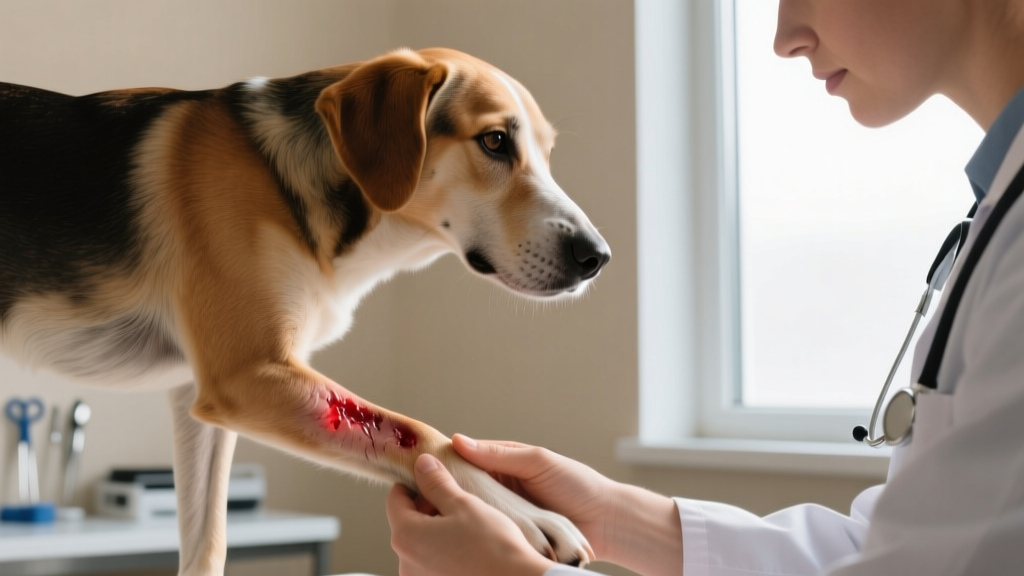
Canine Fatty Tumors: Photos, Identification, and Treatment Guide
Learn to identify canine fatty tumors with photo descriptions, symptoms, diagnosis costs, and treatment options. Expert vet-backed guidance for dog owners.

What Are Canine Fatty Tumors (Lipomas)?
A canine fatty tumor — medically known as a lipoma — is a benign growth composed of mature fat cells (adipocytes) that accumulates beneath the skin or, less commonly, between muscle layers. Lipomas are the most common subcutaneous tumor in dogs, with veterinary dermatologists estimating they appear in up to 1-2% of the general dog population at some point, though prevalence climbs dramatically with age. Middle-aged and senior dogs are disproportionately affected, and certain breeds show strong genetic predisposition.
Most lipomas are soft, movable, well-circumscribed lumps that grow slowly and remain non-cancerous. However, a small subset — called infiltrative lipomas — invade surrounding muscle and connective tissue, while an even rarer malignant form called liposarcoma can metastasize. Because a lipoma can feel identical to a mast cell tumor or soft tissue sarcoma on casual palpation, veterinary evaluation is essential for any new lump.
If you've searched for canine fatty tumors photos hoping to compare what you're seeing on your dog, you're taking the right first step — but visual comparison alone cannot confirm a diagnosis. Only fine-needle aspiration or biopsy can definitively differentiate a lipoma from a potentially dangerous mass.
Recognizing Canine Fatty Tumors: What They Look and Feel Like
Understanding the physical characteristics of lipomas helps you monitor your dog more effectively between vet visits. Here's what to look for:
Typical Appearance and Texture
- Shape: Round to oval, with smooth, well-defined borders
- Texture: Soft and doughy to the touch — similar to pressing on a ripe peach
- Mobility: Moves freely under the skin when gently pushed (non-adherent)
- Skin surface: Overlying skin usually appears normal — no redness, ulceration, or hair loss
- Size range: Typically 0.5 to 3 inches (1.3-7.5 cm) in diameter, though some grow to softball size or larger
- Growth rate: Slow and steady, often enlarging over months to years
- Location: Most commonly found on the chest, abdomen, flanks, and upper legs
Symptom Checklist by Severity
| Severity Level | Symptoms | Action Required |
|---|---|---|
| Early / Mild | Small, soft lump under skin; no pain on palpation; dog shows no behavioral changes; lump present for weeks without rapid growth | Schedule routine vet appointment; measure and photograph for monitoring |
| Moderate | Lump growing noticeably over weeks; multiple new lumps appearing; lump interfering with movement or harness fit; slight firmness developing | Schedule vet visit within 1-2 weeks; discuss fine-needle aspiration |
| Advanced / Concerning | Rapid growth (noticeable change in days); lump feels hard or fixed to underlying tissue; skin discoloration or ulceration over mass; limping or reluctance to move; weight loss or lethargy; lump in armpit or groin impeding limb function | Urgent vet appointment within 24-48 hours; may indicate infiltrative lipoma or different tumor type |
According to the Merck Veterinary Manual, the distinction between a simple lipoma and an infiltrative lipoma is clinically significant — infiltrative lipomas send finger-like projections into muscle fascia and can cause lameness or nerve compression even though they remain technically benign.
Risk Factors: Which Dogs Are Most Prone to Fatty Tumors?
Detailed Risk Factor Breakdown
| Risk Factor | Details | Relative Risk |
|---|---|---|
| Age | Most lipomas appear in dogs 6-12 years old; rare in dogs under 4 | High — age is the strongest predictor |
| Weight / Body Condition | Overweight dogs (BCS 6-7/9) and obese dogs (BCS 8-9/9) develop lipomas at significantly higher rates | High — excess adipose tissue is a direct contributor |
| Breed | Labrador Retrievers, Weimaraners, Dobermans, Schnauzers, Cocker Spaniels, Dachshunds, Pit Bull-type breeds | Moderate to High — genetic component confirmed |
| Sex | Female dogs show slightly higher prevalence, particularly spayed females | Moderate |
| Hormonal Status | Hypothyroidism and Cushing's disease (hyperadrenocorticism) are associated with increased lipoma formation | Moderate — treat underlying endocrine condition |
| Environment | No strong environmental link confirmed; some anecdotal reports suggest pesticide exposure, but evidence is limited | Low / Uncertain |
Why Overweight Dogs Develop More Lipomas
The connection between obesity and lipoma formation is well-documented. Adipose tissue isn't inert storage — it's an active endocrine organ that produces inflammatory cytokines and growth factors. When a dog carries excess body fat, the increased adipocyte activity creates more opportunities for localized fat cell clusters to form benign tumors. Maintaining your dog at an ideal body condition score (BCS) of 4-5 out of 9 is one of the few modifiable risk factors available.
How Veterinarians Diagnose Canine Fatty Tumors
When you bring a lump to your veterinarian, expect a systematic diagnostic process. Here's a step-by-step overview of what to expect and what each step costs:
- Physical Examination ($50-$85 office visit fee): Your vet will palpate the lump — noting size, shape, texture, mobility, and relationship to underlying structures. They'll check for multiple masses and assess overall body condition.
- Fine-Needle Aspiration / Cytology ($25-$75): A small needle extracts cells from the mass. The sample is smeared on a slide and examined under a microscope. A lipoma yields mature adipocytes (fat cells) that are easily identifiable. This is the gold-standard first-line test and is usually performed during the same visit.
- Biopsy or Excisional Biopsy ($200-$500): If FNA results are inconclusive, if the mass feels atypical (firm, fixed, or irregular), or if the vet suspects an infiltrative lipoma or liposarcoma, a tissue sample or the entire mass may be surgically removed and sent to a veterinary pathologist for histopathology.
- Advanced Imaging — Ultrasound or MRI ($300-$1,200): Recommended for large masses, masses near vital structures, or suspected infiltrative lipomas that may extend deeper than palpation suggests. MRI provides the clearest picture of tissue infiltration but is available primarily at veterinary specialty centers.
- Blood Work ($80-$200): A complete blood count (CBC) and chemistry panel may be recommended to assess overall health before any surgical procedure and to screen for underlying endocrine conditions like hypothyroidism or Cushing's disease that can promote lipoma growth.
In most straightforward cases, a physical exam plus fine-needle aspiration is sufficient. Total diagnostic cost for a simple lipoma confirmation typically runs $75-$160. Complex cases requiring imaging and biopsy can range from $500 to $1,700+ depending on your geographic area and whether a specialty center is involved.
Why Fine-Needle Aspiration Isn't Always Enough
FNA has a diagnostic accuracy of approximately 85-90% for lipomas when performed by an experienced veterinarian. However, it can miss:
- Infiltrative lipomas — the aspirate may look identical to a simple lipoma, but the tumor's behavior is invasive
- Liposarcomas — malignant fat cell tumors are rare but may be under-sampled on FNA
- Mixed tumors — a mass that contains both fatty and non-fatty components may yield only fat cells on aspiration, masking a more serious component
This is why your vet may recommend re-evaluation of a previously diagnosed lipoma if it changes in character — growth pattern, firmness, or skin involvement.
Treatment Options for Canine Fatty Tumors
The good news: most simple lipomas require no treatment at all. However, certain situations warrant intervention. Here's a comparison of available approaches:
| Treatment | When Used | Success Rate | Cost Range | Recovery |
|---|---|---|---|---|
| Watchful Waiting (Monitoring) | Small, confirmed lipomas not causing discomfort or mobility issues | N/A — lipoma remains but is tracked | $0 ongoing (vet rechecks $50-$85) | None needed |
| Surgical Excision | Large lipomas, growing lipomas, masses impeding movement, infiltrative lipomas, or owner preference | 95%+ complete cure for simple lipomas; infiltrative lipomas have 30-50% recurrence if margins are incomplete | $200-$800 per mass (includes anesthesia, surgery, pathology) | 10-14 days; E-collar, restricted activity |
| Liposuction | Very large lipomas where full excision would leave a significant wound; cosmetic concerns | Variable — higher recurrence than excision; not all clinics offer | $400-$1,200 | 7-10 days; less invasive |
| Steroid Injection (Triamcinolone) | Some vets use intralesional steroid injections to shrink lipomas; limited evidence base | Inconsistent — some reduction in size reported, but not a standard protocol | $50-$150 per injection | Minimal; possible local skin atrophy |
| Radiation Therapy | Infiltrative lipomas with incomplete surgical margins; not used for simple lipomas | ~70% local control for infiltrative lipomas when combined with surgery | $2,000-$5,000+ (specialty center) | Multiple sessions over 3-4 weeks |
| Weight Management | All overweight dogs with lipomas — foundational supportive care | Existing lipomas rarely shrink with weight loss, but new lipoma formation may decrease | $20-$60/month (prescription weight-loss diet) | Gradual — target 1-2% body weight loss per week |
When Surgery Is Recommended Over Monitoring
Your veterinarian will typically recommend surgical removal in these scenarios:
- The lipoma is growing rapidly (doubling in size within weeks)
- The mass is impeding movement — commonly seen in armpit (axillary) or groin (inguinal) locations
- The lipoma is exceedingly large — masses over 4-5 inches (10-13 cm) can cause skin tension, pressure necrosis, or discomfort
- FNA results are inconclusive or suspicious — the vet wants a definitive histopathology diagnosis
- The lipoma is infiltrative — invading muscle or fascia and causing functional problems
- The dog is young enough that the lipoma will likely continue growing for years, making eventual surgery riskier at an older age
Surgical removal of a simple lipoma is generally straightforward. Because lipomas are encapsulated (surrounded by a thin fibrous capsule), they often "shell out" cleanly. Recovery involves 10-14 days of incision care, an Elizabethan collar, and activity restriction. Complication rates are low — under 5% in healthy dogs — with seroma (fluid accumulation at the surgical site) being the most common post-operative issue.
Prognosis and Life Expectancy
| Lipoma Type | Prognosis | Life Expectancy Impact | Recurrence Risk |
|---|---|---|---|
| Simple Lipoma | Excellent — benign, non-invasive, no metastatic potential | None — does not affect lifespan | Very low after complete excision; new lipomas may develop elsewhere |
| Infiltrative Lipoma | Good to Fair — locally invasive but does not metastasize; can cause significant local tissue damage | Generally normal lifespan if managed; quality of life may be affected if tumor impinges on nerves or joints | 30-50% recurrence after surgery alone; lower with radiation adjunct |
| Liposarcoma (Malignant) | Guarded — rare but aggressive; can metastasize to lungs and other organs | Variable — depends on grade, stage at diagnosis, and surgical margins; median survival times range from 6 months to 2+ years with aggressive treatment | High if incomplete margins; metastasis possible |
The overwhelming majority of fatty tumors — estimated at over 95% of cases — are simple lipomas with an excellent prognosis. Your dog's life expectancy is not reduced by having one or more confirmed simple lipomas. The critical variable is ensuring accurate diagnosis so that the rare but serious liposarcoma is not missed.
Home Care, Monitoring, and Daily Management
If your veterinarian has confirmed a lipoma and recommended watchful waiting, here's how to manage it at home:
Monthly Monitoring Protocol
- Measure: Use a flexible measuring tape or calipers to record the lump's longest dimension. Note measurements in a dedicated journal or phone note.
- Photograph: Take a clear photo with a coin or ruler next to the lump for scale. Consistent lighting and angle help track subtle changes.
- Palpate: Gently feel the lump for changes in texture (soft → firm), mobility (freely movable → fixed), or skin changes (normal → red, warm, ulcerated).
- Count: Note any new lumps that have appeared since the last check. Multiple lipomas are common in predisposed breeds — some Labrador Retrievers develop dozens over their lifetime.
- Record: Keep a running log with dates, measurements, and observations. Share this with your vet at annual exams.
Lifestyle Adjustments
- Weight management: If your dog is overweight, work with your vet to develop a calorie-restricted feeding plan. Target a gradual loss of 1-2% of body weight per week. Prescription weight-management diets like Hill's Metabolic or Royal Canin Satiety can help.
- Exercise: Maintain regular daily exercise — at least 30-60 minutes of moderate activity for most breeds. This supports weight management and overall metabolic health.
- Harness and gear: If a lipoma is located where a harness or collar sits, switch to a different style that doesn't compress the mass. A front-clip harness or a padded vest-style harness can redistribute pressure.
- Sleeping surface: For dogs with large lipomas on their sides or hips, an orthopedic memory-foam bed reduces pressure on the mass and improves comfort.
- A previously soft lipoma becomes firm, hard, or fixed to underlying tissue
- Rapid growth — noticeable size increase within days to a week
- Skin over the lump becomes red, hot, ulcerated, or begins draining fluid
- Your dog begins limping, favoring a limb, or showing signs of pain near the mass
- Your dog becomes lethargic, loses appetite, or loses weight unexpectedly
- A new lump appears that feels different from previously diagnosed lipomas
Any of these changes could indicate an infiltrative lipoma, a different tumor type developing near a known lipoma, or a lipoma that has outgrown its blood supply. Don't wait for the next scheduled recheck — call your veterinarian promptly.
Treatment Costs: What to Budget For
Understanding costs helps you plan financially and avoid delays in care. Here's a realistic breakdown based on 2025-2026 veterinary pricing across US markets:
| Service | Low-End Cost | Average Cost | High-End Cost |
|---|---|---|---|
| Office visit / exam fee | $50 | $70 | $120 |
| Fine-needle aspiration + cytology | $25 | $50 | $75 |
| Surgical excision (per mass, incl. anesthesia & pathology) | $200 | $450 | $800+ |
| Multiple mass removal (3+ lipomas, same session) | $500 | $900 | $1,500+ |
| Pre-surgical blood work (CBC/chemistry) | $80 | $130 | $200 |
| Ultrasound (if needed) | $300 | $450 | $600 |
| MRI (specialty center, if needed) | $800 | $1,200 | $2,000+ |
| Radiation therapy (infiltrative lipoma, full course) | $2,000 | $3,500 | $5,000+ |
Worst-case scenario budgeting: If your dog develops an infiltrative lipoma requiring surgery, advanced imaging, and radiation therapy, total costs can reach $4,000-$7,000+. Pet insurance can help — many comprehensive plans cover tumor removal and oncology treatments, though pre-existing condition exclusions apply. If your dog is prone to lipomas, enrolling in insurance before any masses appear is financially prudent.
Cost-saving tip: If your dog has multiple lipomas that all need removal, ask your vet about removing several during a single anesthetic event. This consolidates anesthesia costs and reduces total expense compared to staging separate surgeries.
Can Fatty Tumors Be Prevented?
There is no guaranteed way to prevent lipomas — genetics and age play dominant roles. However, you can reduce risk and severity through these strategies:
- Maintain ideal weight: This is the single most impactful modifiable factor. Dogs at an ideal BCS (4-5/9) develop fewer lipomas than overweight counterparts. Use measured portions rather than free-feeding, and limit treats to 10% of daily caloric intake.
- Regular veterinary exams: Bi-annual wellness checks for senior dogs (age 7+) allow early detection. Ask your vet to perform a full-body "lump check" at every visit — running hands over the entire body to map all masses.
- Screen for endocrine disease: If your dog is developing multiple lipomas, ask your vet about thyroid function testing (total T4, free T4, TSH) and screening for Cushing's disease (ACTH stimulation test or low-dose dexamethasone suppression test). Treating an underlying hormonal imbalance may slow new lipoma formation.
- Avoid unnecessary supplements marketed as "lipoma cures": No supplement has been scientifically proven to prevent or dissolve canine lipomas. Products claiming to do so are not FDA-approved and may contain undisclosed ingredients. Always consult your veterinarian before starting any supplement.
Frequently Asked Questions
Can I tell if my dog's lump is a fatty tumor just by looking at photos online?
No. While canine fatty tumors photos can help you understand what lipomas typically look like — soft, round, under-the-skin masses — visual identification is unreliable. Mast cell tumors, soft tissue sarcomas, and other serious growths can look and feel remarkably similar to lipomas. Only a veterinarian performing fine-needle aspiration or biopsy can provide a definitive diagnosis. Use online photos as educational context, not as a diagnostic tool.
Do fatty tumors go away on their own?
No. Once a lipoma forms, it does not spontaneously resolve. It may remain the same size for years, or it may continue to grow slowly. Weight loss can sometimes reduce the size of a lipoma slightly (since it's composed of fat cells that can shrink), but the tumor structure itself will not disappear without surgical removal.
My dog has multiple fatty tumors — should I have them all removed?
Not necessarily. Most veterinarians recommend removing lipomas only if they are growing rapidly, causing discomfort, impeding movement, or if the diagnosis is uncertain. Dogs with a genetic tendency toward lipomas — especially Labrador Retrievers — may develop 10, 20, or even more lipomas over their lifetime. Removing all of them would subject the dog to repeated surgeries with cumulative anesthesia risk. A practical approach is to FNA each new lump for confirmation, then remove only those that are problematic.
At what age do dogs typically get fatty tumors?
Lipomas are most commonly diagnosed in dogs between 6 and 12 years of age. They are uncommon in dogs under 4 years old. If a young dog develops a fatty mass, your vet may want to investigate more carefully, as lipomas in young dogs are less typical and other tumor types should be considered.
Are fatty tumors cancerous?
Simple lipomas are not cancerous — they are benign growths that do not metastasize or invade surrounding tissues. However, two related conditions exist on a spectrum: infiltrative lipomas (locally invasive but non-metastatic) and liposarcomas (malignant and capable of spreading). This is why professional diagnosis matters — you cannot distinguish these by feel alone.
How much does it cost to remove a fatty tumor from a dog?
Surgical removal of a single simple lipoma typically costs $200-$800 in the United States, depending on the mass size, location, your geographic area, and whether the procedure is performed at a general practice or specialty center. This includes the exam, pre-surgical bloodwork, anesthesia, surgery, and histopathology. Larger or more complex masses cost more.
What breeds are most prone to developing fatty tumors?
According to the American Kennel Club and veterinary dermatology literature, the breeds most commonly affected include Labrador Retrievers, Weimaraners, Doberman Pinschers, Miniature Schnauzers, Cocker Spaniels, Dachshunds, and Pit Bull-type breeds. Mixed-breed dogs are also commonly affected, particularly those that are overweight or senior.
Should I be worried if my dog's fatty tumor is growing?
Slow, gradual growth over months to years is typical for simple lipomas and is not inherently alarming. However, rapid growth — noticeable size changes within days or weeks — is a red flag that warrants prompt veterinary evaluation. Rapid growth can indicate an infiltrative lipoma, a liposarcoma, or a completely different tumor type that was misdiagnosed. Consult your veterinarian for re-evaluation any time a known lipoma changes character.
Can diet or supplements shrink my dog's fatty tumor?
There is no scientifically validated diet or supplement that dissolves or shrinks canine lipomas. While weight loss may slightly reduce the size of fat-based tumors, the tumor capsule and structure remain. Be cautious of online products claiming to "melt away" lipomas — these are unregulated and potentially unsafe. Always discuss dietary changes and supplements with your veterinarian.
This article is for informational purposes only and does not replace professional veterinary advice. If your dog has a new or changing lump, consult your veterinarian for proper diagnosis and treatment. Every mass should be individually evaluated — what looks like a simple fatty tumor may require testing to confirm.
Tom Renshaw
All our authors care for dogs every day — read more of their work on the authors page.