Causes, Symptoms, and What Owners Need to Know
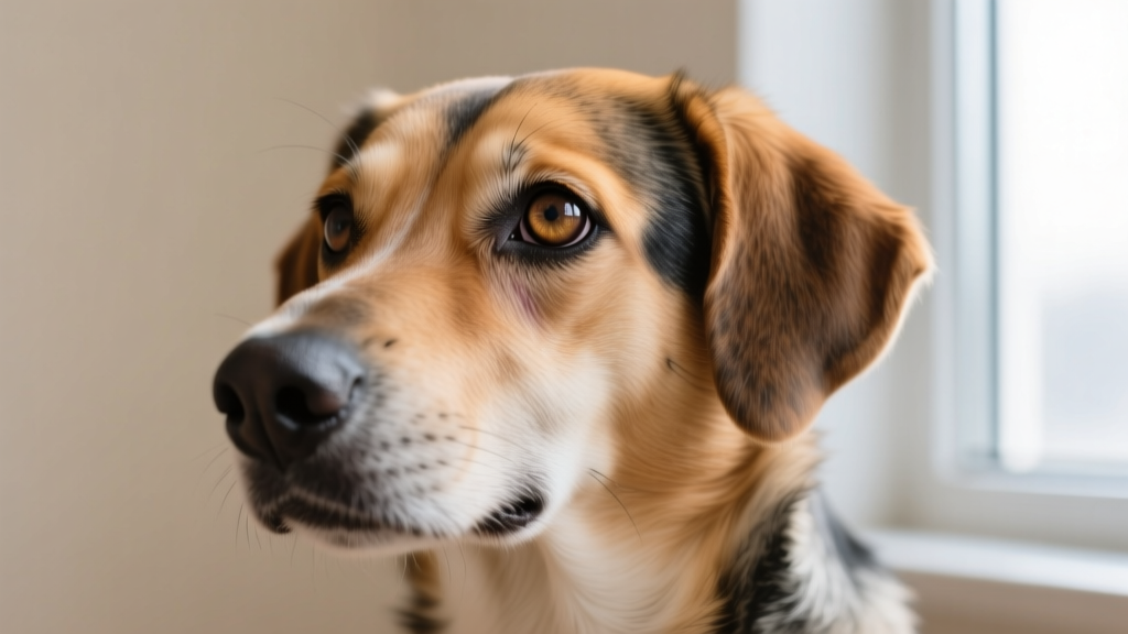
Horner's syndrome in dogs causes a droopy eye, sunken appearance, and small pupil.
Quick Answer: What Is Horner's Syndrome in Dogs?
Horner's syndrome in dogs is a neurological disorder caused by disruption of the sympathetic nerve pathway to the eye. It produces a characteristic cluster of signs on one side of the face: a drooping upper eyelid (ptosis), a constricted pupil (miosis), a sunken-looking eye (enophthalmos), and a protruding third eyelid. In most dogs, Horner's syndrome is idiopathic — meaning no specific cause is found — and it often resolves on its own within 6 to 8 weeks. However, because it can sometimes signal an underlying injury, tumor, or middle ear disease, a veterinary exam is essential to rule out serious conditions.
If you've noticed your dog's eye looking "off" — one pupil smaller than the other, an eyelid that seems to sag, or a strange membrane creeping across the corner of the eye — you're likely dealing with Horner's syndrome. It looks alarming, and the first question most owners ask is whether it's an emergency. The answer depends on what's causing it.
This guide breaks down the science behind Horner's syndrome in dogs: the neuroanatomy involved, why it happens, what your veterinarian will look for, and what you can expect during recovery. Understanding the sympathetic nervous system pathway makes the symptoms far less mysterious and helps you advocate for your dog during diagnosis.
The Sympathetic Nerve Pathway: How Horner's Syndrome Develops
The Three-Neuron Chain to the Eye
The sympathetic innervation of the eye follows a remarkably long and vulnerable pathway — a three-neuron chain that travels from the brain, down the spinal cord, back up through the neck, and into the face. Damage at any point along this chain can produce Horner's syndrome.
First-order neuron (central): Originates in the hypothalamus and descends through the brainstem and cervical spinal cord. Lesions here are rare but can result from brain tumors, strokes, or spinal cord trauma in the neck region.
Second-order neuron (preganglionic): Exits the spinal cord at the level of the first three thoracic vertebrae (T1–T3), travels through the chest cavity, and ascends the neck via the vagosympathetic trunk. This segment is vulnerable to chest tumors (especially anterior mediastinal lymphoma), neck trauma, and surgical complications from procedures near the jugular vein or thyroid.
Third-order neuron (postganglionic): Synapses at the cranial cervical ganglion near the base of the skull, then travels along the internal carotid artery through the middle ear and into the orbit of the eye. This is the most commonly affected segment in dogs — middle ear disease (otitis media), nasopharyngeal polyps, and trauma to the retrobulbar area are frequent culprits.
According to the Merck Veterinary Manual, the postganglionic (third-order) pathway is the most frequently implicated in canine Horner's syndrome cases. The nerve fibers at this stage are thin, unmyelinated, and travel through tight anatomical spaces — making them especially susceptible to compression, inflammation, and mechanical irritation.
What Happens When Sympathetic Input Is Lost
The sympathetic nervous system normally maintains several structures in the eye and face in a state of constant low-level activation (sympathetic tone). When that input is disrupted, the opposing parasympathetic system dominates, producing the four classic clinical signs:
| Clinical Sign | Mechanism | What You See |
|---|---|---|
| Ptosis | Loss of sympathetic stimulation to the superior tarsal (Müller's) muscle in the upper eyelid | Upper eyelid droops, partially covering the eye |
| Miosis | Unopposed parasympathetic constriction of the iris sphincter muscle (pupillary dilator loses sympathetic drive) | Affected pupil is noticeably smaller than the normal eye |
| Enophthalmos | Loss of sympathetic tone to the orbitalis muscle (smooth muscle behind the globe) | Eye appears sunken deeper into the socket |
| Third eyelid protrusion | Passive result of enophthalmos — as the globe recedes, the nictitating membrane becomes more visible | Pink or pale membrane visible at the inner corner of the eye |
Some dogs also show facial warmth and vasodilation on the affected side (loss of sympathetic vasoconstriction), though this is harder to observe in dogs than in humans and is not always clinically apparent.
Common Causes of Horner's Syndrome in Dogs
Understanding the likely causes helps you and your veterinarian narrow the diagnostic focus. Causes are generally categorized by which neuron in the three-neuron chain is affected:
Idiopathic (No Identified Cause)
In approximately 40–50% of canine cases, no underlying cause is ever identified. Idiopathic Horner's syndrome is a diagnosis of exclusion — your vet rules out trauma, tumors, ear disease, and other conditions, and the syndrome resolves spontaneously. Studies published in the Journal of Veterinary Internal Medicine suggest idiopathic cases tend to have a faster recovery (often 6–8 weeks) compared to those with identifiable causes.
Trauma
Blunt trauma to the head, neck, or chest can damage any segment of the sympathetic pathway. Common scenarios include:
- Bite wounds to the neck area
- Hit-by-car injuries affecting the thoracic inlet or cervical spine
- Aggressive leash-pulling injuries (especially in dogs wearing choke or slip collars — another reason to use a well-fitted harness)
- Iatrogenic (post-surgical) trauma from procedures involving the neck, chest, or ear canal
Otitis Media (Middle Ear Infection)
The postganglionic sympathetic fibers pass directly through or adjacent to the middle ear cavity. Chronic or acute otitis media — particularly in breeds prone to ear infections — can compress or inflame these fibers. This is one of the more treatable identifiable causes, as resolving the ear infection often allows nerve recovery.
Neoplasia (Tumors)
Tumors along the sympathetic pathway are a serious but less common cause. These include:
- Anterior mediastinal lymphoma or thymoma (affecting the preganglionic neuron in the chest)
- Nerve sheath tumors along the vagosympathetic trunk
- Nasopharyngeal polyps or tumors near the cranial cervical ganglion
- Retrobulbar masses behind the eye
Breed Predispositions
While Horner's syndrome can occur in any breed, certain breeds appear overrepresented in veterinary literature:
| Breed | Noted Pattern |
|---|---|
| Golden Retriever | Most commonly reported breed in idiopathic cases; multiple retrospective studies show overrepresentation |
| Cocker Spaniel | Higher rates of otitis media may contribute to increased secondary Horner's cases |
| Collie / Shetland Sheepdog | Reported in idiopathic clusters; possible genetic predisposition under investigation |
| Dachshund | IVDD-related spinal cord issues may occasionally involve first-order neuron pathways |
Golden Retrievers, in particular, appear in veterinary literature with idiopathic Horner's syndrome at a rate that suggests a possible breed-specific susceptibility, though no single gene has been identified. If you own a Golden Retriever and notice these eye changes, idiopathic Horner's is statistically the most likely diagnosis — but your vet will still want to rule out other causes.
How Veterinarians Diagnose Horner's Syndrome
Diagnosis begins with recognizing the classic tetrad of clinical signs. However, your veterinarian will perform additional tests to determine where along the pathway the disruption has occurred and why.
Pharmacologic Localization (Eye Drop Testing)
This is the most valuable diagnostic tool for pinpointing the lesion site. Two agents are used:
How the Eye Drop Test Works
Step 1 — Hydroxyamphetamine (1%): This drug causes the release of stored norepinephrine from intact postganglionic nerve terminals. If the third-order (postganglionic) neuron is damaged, there are no intact terminals to release norepinephrine, and the pupil will not dilate. If the lesion is first- or second-order, the pupil will dilate because the postganglionic neuron is still intact.
Step 2 — Phenylephrine (1% or 2.5%): This directly stimulates the iris dilator muscle (bypassing the nerve entirely). In a normal eye, dilation takes 30–40 minutes. In a denervated eye (postganglionic lesion), the iris dilator muscle becomes hypersensitive due to denervation upregulation — the pupil dilates in under 20 minutes. If the lesion is preganglionic or central, dilation timing is normal.
Interpretation:
- Rapid dilation with phenylephrine (<20 min) + no dilation with hydroxyamphetamine = third-order (postganglionic) lesion
- Normal dilation timing with both = first- or second-order lesion (further imaging needed)
Note: Hydroxyamphetamine can be difficult to source in veterinary practice. Many clinicians rely primarily on phenylephrine testing combined with imaging and clinical history.
Additional Diagnostics
Depending on the suspected lesion location, your vet may recommend:
- Otoscopic examination — to check for middle ear disease, polyps, or masses in the ear canal
- Advanced imaging (CT or MRI) — typically $1,200–$2,500 depending on your region and facility. MRI is preferred for evaluating the brain, brainstem, and cervical spinal cord. CT is better for assessing the middle ear and bony structures of the skull base
- Chest radiographs or CT — to rule out mediastinal masses if a preganglionic lesion is suspected
- Complete neurological exam — to assess for other cranial nerve deficits, proprioception abnormalities, or spinal pain that might indicate a central lesion
- Schirmer tear test — because sympathetic disruption can occasionally reduce tear production on the affected side
What to Expect at the Vet Visit
- Initial exam + phenylephrine test: $80–$200
- Otoscopic exam: Often included in the exam fee; deep ear cleaning under sedation adds $150–$350
- MRI (brain/cervical spine): $1,500–$2,500
- CT scan: $1,200–$2,000
- Chest X-rays (3-view): $200–$400
Total diagnostic costs can range from $80 (idiopathic case with minimal workup) to $3,000+ (full imaging panel). Pet insurance with illness coverage can significantly offset these costs if you enrolled before symptom onset.
Treatment Options and Recovery Timeline
Treatment depends entirely on the underlying cause. There is no specific "cure" for Horner's syndrome itself — the goal is to address the root problem and allow the nerve to heal.
Idiopathic Cases
No treatment is required beyond monitoring. Phenylephrine eye drops (2.5%) may be prescribed for cosmetic improvement — they temporarily dilate the pupil and reduce the droopy appearance for a few hours. This is purely symptomatic and does not speed recovery. Most idiopathic cases resolve within 6 to 8 weeks, though some take up to 4 months.
Otitis Media
Aggressive treatment of the ear infection — typically systemic antibiotics (based on culture and sensitivity results) for 4–6 weeks, plus topical ear medications and possibly ear flushing under anesthesia. Horner's signs may resolve within weeks of infection control, though nerve recovery can lag behind clinical improvement by several weeks.
Trauma
Supportive care and time. Anti-inflammatory medications (such as a short course of corticosteroids or NSAIDs, as directed by your vet) may help reduce nerve inflammation. Nerve regeneration occurs at approximately 1–2 mm per day, so recovery depends on the extent and location of damage. Some traumatic cases result in permanent partial Horner's syndrome.
Neoplasia
Treatment depends on the tumor type, location, and staging. Options include surgical removal, chemotherapy (for lymphoma), or radiation therapy. Prognosis varies widely — mediastinal lymphoma in dogs has a median survival time of approximately 6–12 months with chemotherapy, though individual outcomes depend on the stage and the dog's overall health.
When to See the Vet Immediately
Contact your veterinarian or an emergency clinic if your dog shows Horner's syndrome signs alongside any of the following:
- Head tilt, circling, or loss of balance (suggests central nervous system involvement)
- Difficulty swallowing or voice changes
- Weakness or incoordination in the limbs
- Signs of pain — vocalizing, reluctance to move, neck guarding
- Horner's signs appearing after recent trauma (car accident, fall, fight)
- Rapid onset of additional neurological symptoms
Isolated Horner's syndrome without other symptoms is generally not an emergency, but should still be evaluated within 24–48 hours.
Evolutionary Context: Why the Sympathetic Eye Pathway Exists
From Ancestral Predators to Modern Dogs
The sympathetic innervation of the eye is not unique to dogs — it's an ancient vertebrate system shared across mammals, birds, and reptiles. In wolves and other ancestral canids, this system served critical survival functions:
Pupil dilation for threat assessment: Sympathetic activation rapidly dilates the pupils during fight-or-flight responses, maximizing light intake and widening the visual field. For a predator scanning for prey or assessing a rival pack, this split-second visual enhancement could mean the difference between eating and going hungry.
Eyelid retraction for visual clarity: The superior tarsal muscle (Müller's muscle) elevates the upper eyelid beyond what the skeletal levator muscle alone can achieve. This provides a wider, unobstructed visual field — critical for an animal that relies on detecting movement across open terrain.
Orbital muscle and eye protection: The orbitalis smooth muscle, innervated sympathetically, positions the globe optimally within the orbit. During high-stress encounters, sympathetic activation pushes the eye slightly forward, improving the field of view. When the animal is calm, the eye sits slightly deeper, offering more bony protection.
The vulnerability of this pathway — its long, exposed route through the neck and chest — is an evolutionary trade-off. The sympathetic chain had to traverse these regions to reach the head from the thoracic spinal cord, and there was no selective pressure to "reroute" it because the pathway worked well enough for survival. In modern domestic dogs, this same anatomical layout persists, and the pathway remains susceptible to the same disruptions that would have affected wolves — trauma to the neck, infections spreading from the ear, and masses along the nerve course.
Common Misconceptions About Horner's Syndrome in Dogs
| Myth | Fact |
|---|---|
| "Horner's syndrome means my dog has a brain tumor." | Most cases are idiopathic or related to ear disease. Brain tumors are a rare cause, and they typically produce additional neurological symptoms beyond the eye signs. |
| "It's painful for my dog." | Horner's syndrome itself is not painful. The eye changes are neurological, not inflammatory. However, an underlying cause (ear infection, trauma) may cause discomfort. |
| "My dog will go blind from this." | Horner's syndrome does not cause blindness. Vision is typically unaffected, though the drooping eyelid may partially obstruct the visual field until resolved. |
| "It's contagious to other dogs or to me." | Horner's syndrome is a neurological disruption, not an infection. It cannot spread between animals or to humans. |
| "Eye drops will cure it." | Phenylephrine drops are a diagnostic tool and cosmetic aid — they temporarily mask symptoms but do not treat the underlying nerve disruption. |
| "If it doesn't go away quickly, something is seriously wrong." | Even idiopathic cases can take 2–4 months to fully resolve. Slow improvement doesn't necessarily indicate a worse prognosis. |
How Horner's Syndrome Differs in Dogs vs. Humans
While the neurological mechanism is essentially the same across mammals, there are practical differences worth understanding:
- Anhidrosis (loss of facial sweating) is a classic sign in human Horner's syndrome but is not clinically observable in dogs, since dogs do not rely on facial sweat glands for thermoregulation. Their sweat glands are concentrated in the paw pads.
- Diagnostic pharmacology differs: The same eye drop tests are used, but dogs may require higher phenylephrine concentrations or slightly longer observation windows due to species-specific iris muscle sensitivity.
- Idiopathic rates are higher in dogs: In human medicine, Horner's syndrome more frequently has an identifiable structural cause (carotid dissection, Pancoast tumors, surgical complications). Dogs have a higher proportion of idiopathic cases, which is somewhat reassuring from a prognostic standpoint.
- Communication barrier: Humans with Horner's syndrome can describe visual disturbances, headaches, or neck pain that help localize the lesion. Dogs cannot, making imaging and pharmacologic testing even more critical for accurate diagnosis.
Practical Implications for Daily Life
What Changes (and What Doesn't) When Your Dog Has Horner's Syndrome
Exercise and activity: Unless your vet has identified a cause that requires rest (spinal injury, post-surgical recovery), dogs with idiopathic Horner's syndrome can continue normal exercise. Their vision and physical ability are not meaningfully impaired.
Eye care: The protruding third eyelid and slight eyelid droop can reduce the eye's natural ability to distribute tears evenly. Monitor for signs of dryness — excessive blinking, squinting, or mucoid discharge. Your vet may recommend a lubricating eye gel (such as Genteal or a veterinary-specific product like I-DROP VET) applied 2–3 times daily as a preventive measure. These gels typically cost $12–$25 per tube and last several weeks.
Monitoring at home: Check the affected eye daily for:
- Increasing redness or discharge (possible secondary conjunctivitis or corneal ulcer from inadequate eyelid coverage)
- Signs of pain — pawing at the eye, rubbing the face on furniture
- Changes in pupil size — if the affected pupil begins to dilate or if the normal eye changes, contact your vet
- Any new neurological signs — head tilt, balance issues, changes in eating or drinking behavior
Grooming considerations: Keep hair trimmed around the affected eye to prevent irritation from the drooping lid. Avoid getting shampoo or grooming products in the eye, as reduced blink efficiency may slow clearance of irritants.
When to recheck: If your dog was diagnosed with idiopathic Horner's syndrome, schedule a recheck at 4 weeks and again at 8 weeks. If there is no improvement by 8 weeks, your vet may recommend advanced imaging even if the initial workup was normal.
Related Conditions to Be Aware Of
Understanding conditions that can mimic or accompany Horner's syndrome helps you communicate more effectively with your veterinarian:
- Facial nerve paralysis: Can cause a drooping face and eyelid but does NOT produce miosis (small pupil). The pupil remains normal. Often seen in older Cocker Spaniels and may be idiopathic or related to hypothyroidism.
- Uveitis: Inflammation inside the eye can cause a constricted pupil and third eyelid elevation, but is accompanied by significant redness, cloudiness, and pain. Uveitis is an ophthalmologic emergency requiring immediate treatment to prevent glaucoma or blindness.
- Haw syndrome: Bilateral protrusion of the third eyelids, often associated with gastrointestinal illness. Unlike Horner's, it affects both eyes and does not involve miosis or ptosis. Common in young dogs recovering from diarrhea or parasitic infections.
- Cerebellar or brainstem lesions: Can produce Horner's syndrome along with other cranial nerve deficits, ataxia, and altered mentation. These cases present with obvious additional neurological abnormalities.
Frequently Asked Questions
Why does my dog have a droopy eye and small pupil on one side?
This combination is the hallmark of Horner's syndrome — a disruption of the sympathetic nerve pathway to the eye. The sympathetic nervous system normally keeps the upper eyelid elevated and the pupil dilated. When that input is lost, the eyelid droops (ptosis) and the pupil constricts (miosis) due to unopposed parasympathetic activity. The cause ranges from idiopathic (most common) to ear infections, trauma, or, rarely, tumors.
Can dogs fully recover from Horner's syndrome?
Yes. In idiopathic cases, full recovery occurs in the majority of dogs within 6 to 8 weeks, though some cases take up to 4 months. Recovery depends on the underlying cause — trauma-related cases may have partial permanent deficits, and tumor-related cases depend on successful treatment of the neoplasia. Nerve regeneration is slow but often complete in non-neoplastic cases.
How do dogs process visual information with Horner's syndrome?
Dogs with Horner's syndrome retain normal visual processing — the retina, optic nerve, and visual cortex are all unaffected. The only visual impact is a mildly reduced visual field due to the drooping eyelid, similar to a human looking through half-closed eyes. Once the eyelid returns to normal position, vision is fully restored.
What does it mean when my dog's third eyelid is showing?
A visible third eyelid (nictitating membrane) can indicate Horner's syndrome, but also haw syndrome, dehydration, weight loss (loss of retrobulbar fat), or a mass behind the eye. In Horner's, the third eyelid protrudes passively because the eyeball sits deeper in the socket (enophthalmos). It's not actively "pushing" forward — the globe has receded, revealing the membrane.
Is Horner's syndrome normal or should I panic?
Don't panic, but do schedule a veterinary exam within 24–48 hours. Isolated Horner's syndrome without other neurological symptoms is usually not an emergency, and in many cases, it's idiopathic and self-resolving. However, it should never be ignored because identifying an underlying cause early — especially middle ear disease or a mass — improves treatment outcomes significantly.
Is Horner's syndrome in dogs different from humans?
The core neurological mechanism is identical — disruption of the sympathetic pathway to the eye produces the same signs in both species. The main differences are that dogs don't show facial sweating changes (anhidrosis), idiopathic cases are more common in dogs, and dogs can't describe associated symptoms like pain or visual changes, making veterinary examination and testing essential for proper diagnosis.
Disclaimer: This article is for educational purposes and does not replace professional veterinary advice. If your dog shows signs of Horner's syndrome, consult your veterinarian for a proper diagnosis and treatment plan. References: Merck Veterinary Manual — Horner's Syndrome in Animals, AVMA Pet Owner Resources.
Dr. Hannah Wickes
All our authors care for dogs every day — read more of their work on the authors page.


