Heterochromia Iridum in Dogs: The Science Behind Two-Toned Eyes
What causes heterochromia iridum in dogs? Explore the genetics, melanin science, breed predispositions, and health implications of two-colored eyes in canines.
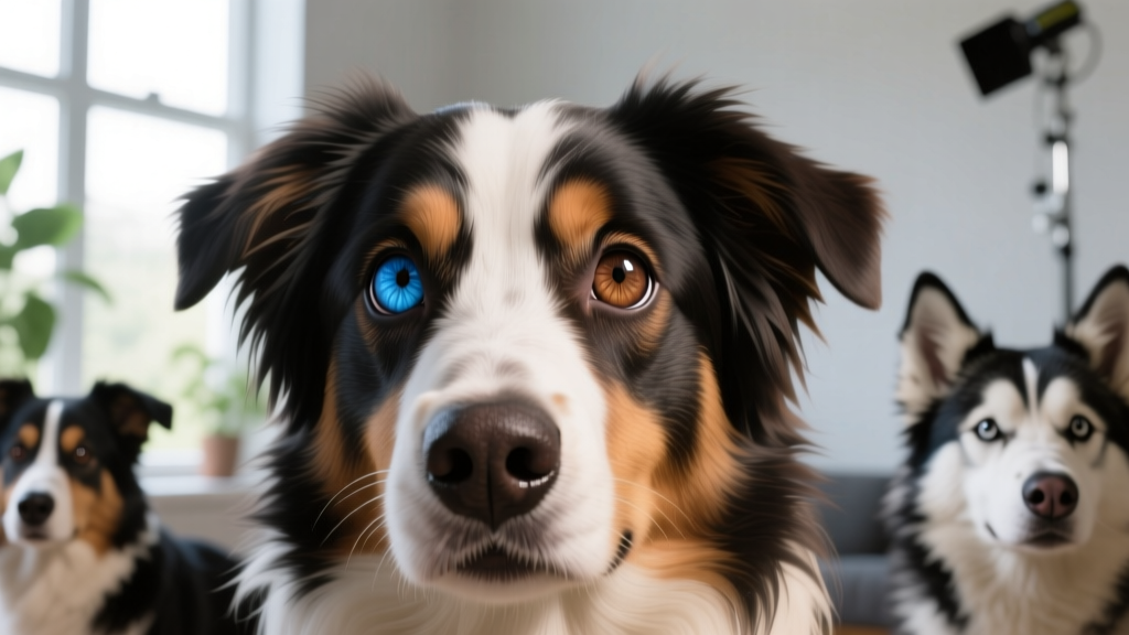
Quick Answer: What Is Heterochromia Iridum in Dogs?
Heterochromia iridum in dogs is a condition where a dog has two differently colored irises (complete heterochromia), multiple colors within a single iris (partial or sectoral heterochromia), or a concentric ring of a different color around the pupil (central heterochromia). It is caused by uneven melanin distribution in the iris stroma during embryonic development, most commonly linked to the merle gene (SILV/PMEL17), piebald spotting (MITF gene), and extreme white coat patterns. In the vast majority of cases, heterochromia is a benign cosmetic trait that does not affect vision or eye health. However, when accompanied by a white coat and blue eyes in certain breeds, it can co-occur with congenital deafness — warranting a BAER hearing test.
If your dog gazes back at you with one icy blue eye and one warm brown, you've likely fielded plenty of questions at the dog park. Heterochromia iridum in dogs is one of the most visually striking traits in the canine world, and it's far more than a quirky accident. It's a window into pigment biology, coat-color genetics, and the evolutionary history of domesticated dogs. Let's unpack what the science actually says.
The Genetics and Melanin Science Behind Heterochromia Iridum
Physiological Mechanism: How the Body Produces Two-Colored Eyes
Eye color in dogs — as in humans — is determined by the concentration and distribution of melanin in the iris stroma (the connective tissue layer of the iris). Melanin is produced by specialized cells called melanocytes, which migrate from the neural crest during embryonic development.
The process works like this:
- High melanin concentration in the iris stroma absorbs more light, producing brown or amber eyes.
- Low melanin concentration allows light to scatter through the stroma (a phenomenon called Tyndall scattering), producing the appearance of blue — even though no blue pigment exists.
- Uneven melanin distribution across the two irises results in heterochromia. One eye may receive a full complement of melanocytes while the other receives fewer, or melanocytes may distribute unevenly within a single iris.
The critical developmental window occurs between approximately days 28–45 of gestation, when neural crest cells migrate to the developing eye. Any genetic factor that disrupts melanocyte migration, survival, or melanin synthesis can result in heterochromia. Puppies are typically born with blue eyes because melanin production in the iris hasn't fully begun; permanent eye color usually establishes between 8 and 16 weeks of age.
The Key Genes Involved
Research published in the National Institutes of Health (NIH) and studies referenced by the Merck Veterinary Manual identify several gene loci strongly associated with heterochromia:
| Gene / Locus | Mechanism | Associated Breeds | Heterochromia Frequency |
|---|---|---|---|
| Merle (M locus / SILV-PMEL17) | Dilutes eumelanin in random patches, affecting coat and iris melanocytes | Australian Shepherd, Border Collie, Great Dane, Dachshund | Very high in merles — estimated 50–80% of merle dogs show some heterochromia |
| Piebald (S locus / MITF gene) | Disrupts melanocyte migration, creating white patches and reducing iris pigment | Dalmatian, Bull Terrier, English Setter, Foxhound | Common in extreme piebald (sp) — roughly 30–50% |
| Extreme White (sw allele) | Near-total suppression of melanocyte migration to skin and iris | Dalmatian, Boxer (white), Dogo Argentino | High — blue or mismatched eyes frequent |
| Alaskan Husky / Northern Breed lineage | Polygenic — multiple genes influence melanin deposition; not solely merle-driven | Siberian Husky, Alaskan Malamute, Alaskan Husky | Siberian Huskies: approximately 40–60% show heterochromia or parti-colored eyes |
A critical distinction: the merle gene (M) is an incomplete dominant. A single copy (Mm) produces the merle pattern with associated heterochromia. Two copies (MM — "double merle") cause severe pigment depletion, frequently resulting in predominantly white coats, bilateral blue eyes, and significantly elevated risks of deafness and ocular abnormalities including microphthalmia and coloboma. Responsible breeders never intentionally produce double-merle pairings.
Evolutionary Context: Why Heterochromia Exists in Canids
Evolutionary Basis
Wild wolves (Canis lupus) almost never display heterochromia. Wolf eyes are consistently amber to yellow-brown, optimized for crepuscular (dawn/dusk) hunting. The trait is overwhelmingly a product of artificial selection during domestication, not natural selection.
Here's the evolutionary logic: as early humans selected dogs for specific coat patterns — white spotting for visibility during hunting, merle patterning for aesthetic preference — the genes controlling melanocyte migration were inadvertently co-selected. The same neural crest cells that produce white coat patches also populate the iris. Selecting for "more white" or "diluted pigment" in the coat necessarily reduces melanin in the eyes.
This is a classic example of pleiotropy — a single gene affecting multiple, seemingly unrelated traits. The MITF gene, for instance, regulates both coat spotting patterns and inner ear melanocyte development (relevant to hearing). Breeders selecting for a white blaze on the face were simultaneously selecting for reduced iris pigmentation and, in some cases, altered auditory function.
Siberian Huskies present an interesting partial exception. Their heterochromia appears linked to a distinct genetic mechanism not fully shared with piebald or merle breeds. A 2018 study published in PLOS Genetics identified a duplication on canine chromosome 18 (near the ALX4 gene) strongly associated with blue eyes in Siberian Huskies, suggesting this breed evolved a separate genetic pathway to reduced iris melanin — one that likely doesn't carry the same deafness risk as piebald-associated blue eyes.
Types of Heterochromia Iridum in Dogs: A Classification Guide
Veterinary ophthalmologists categorize heterochromia into three distinct types, each with different visual presentations:
- Complete heterochromia (heterochromia iridis): Each eye is a fully different color — for example, one brown and one blue. This is the most recognizable form and the one most people picture.
- Sectoral (partial) heterochromia: A single iris contains two or more distinct color zones. A brown iris may have a wedge-shaped blue sector, or a blue iris may contain a brown "slice." This is especially common in merle Australian Shepherds and Border Collies.
- Central heterochromia: A ring of one color surrounds the pupil, with a different color in the outer iris. For instance, a gold ring around the pupil transitioning to green or brown at the periphery. This is subtler and often overlooked.
Some dogs exhibit bilateral sectoral heterochromia — both eyes have multi-colored irises — which is particularly common in merle-patterned breeds and Siberian Huskies. The American Kennel Club (AKC) notes that several breed standards explicitly permit or even celebrate heterochromia, including the Australian Shepherd, Siberian Husky, and Catahoula Leopard Dog.
Breed Variations: Which Dogs Are Most Likely to Have Heterochromia?
| Breed | Primary Genetic Driver | Typical Eye Color Combinations | Deafness Association |
|---|---|---|---|
| Siberian Husky | ALX4 gene duplication (Chr 18) | Blue/brown, blue/green, parti-colored | Low — not strongly linked to congenital deafness |
| Australian Shepherd | Merle (M locus) | Blue/brown, amber/blue, sectoral patterns | Moderate in merles — BAER testing recommended |
| Border Collie | Merle (M locus) | Blue/brown, sectoral blue in brown iris | Moderate in merles |
| Dalmatian | Extreme piebald (sw) | Blue/brown, bilateral blue | High — ~8% unilateral deafness, ~5% bilateral in blue-eyed Dals |
| Great Dane (merle/harlequin) | Merle + Harlequin modifier | Blue/brown, walleye (light iris) | Moderate — double merle risk significant |
| Catahoula Leopard Dog | Merle (M locus) | "Glass eyes" (blue), multi-colored, sectoral | Moderate in double merle lines |
| Shetland Sheepdog | Merle (M locus) | Blue/brown in blue merles | Moderate in merles |
When Heterochromia Might Signal a Health Problem
⚠️ When to Consult Your Veterinarian
While congenital (present from puppyhood) heterochromia is almost always benign, acquired heterochromia — a change in eye color in an adult dog — should always be evaluated by a veterinarian promptly. Potential causes include:
- Uveitis (intraocular inflammation): Chronic inflammation can alter iris pigmentation. Look for squinting, redness, cloudiness, or pawing at the eye.
- Iris melanoma: A dark spot or progressive darkening of one iris sector may indicate a melanocytic tumor. While most canine iris melanomas are benign, they can cause secondary glaucoma.
- Iris atrophy: Age-related thinning of the iris can make one eye appear lighter. Common in senior dogs, especially small breeds. The pupil may appear irregularly shaped.
- Glaucoma: Elevated intraocular pressure can cause corneal clouding that alters apparent eye color. This is a veterinary emergency — irreversible vision loss can occur within 24–72 hours without treatment.
- Horner's syndrome: A neurological condition causing a constricted pupil, drooping eyelid, and apparent color change due to the smaller pupil size. Usually resolves on its own but warrants diagnosis.
Red flags requiring immediate veterinary attention: sudden eye color change, visible mass on the iris, cloudiness or opacity, squinting or blepharospasm, excessive tearing or discharge, behavioral changes suggesting vision loss (bumping into objects, reluctance to navigate stairs).
The Deafness Connection: What Owners of White-Coated, Blue-Eyed Dogs Need to Know
The link between heterochromia, white coat patterns, and congenital sensorineural deafness is well-established. The mechanism: the same melanocytes that pigment the iris also populate the stria vascularis of the inner ear. This structure is essential for maintaining the potassium-rich endolymph that enables cochlear hair cells to transmit sound signals. Without melanocytes in the stria vascularis, hair cells degenerate within the first few weeks of life, resulting in permanent deafness.
Prevalence data by breed and eye color:
- Dalmatians with bilateral blue eyes: approximately 30–40% are unilaterally or bilaterally deaf, compared to ~15% in brown-eyed Dalmatians (source: Strain, 2004, Veterinary Journal).
- White Boxers: estimated 18% unilateral or bilateral deafness in predominantly white dogs.
- Double-merle dogs (any breed): deafness prevalence ranges from 25–50% depending on the extent of pigment loss.
- Siberian Huskies: notably, blue eyes in this breed are not strongly associated with deafness, likely because the ALX4 mechanism doesn't disrupt inner ear melanocytes the same way piebald does.
If you own a dog with heterochromia from a breed with known deafness associations, request a BAER (Brainstem Auditory Evoked Response) test. This non-invasive test measures electrical activity in the auditory pathway and can definitively diagnose unilateral or bilateral deafness. Cost typically ranges from $75–$150 at veterinary teaching hospitals or specialty clinics. Puppies can be tested as early as 5–6 weeks of age.
Common Misconceptions About Heterochromia in Dogs
| Myth | Fact |
|---|---|
| "Dogs with heterochromia are blind or have poor vision in the blue eye." | The blue eye has normal vision. The lack of melanin affects iris color, not the retina or lens. Blue-eyed dogs see just as well as brown-eyed dogs in the affected eye, though very light irises may be slightly more sensitive to bright sunlight. |
| "Heterochromia always means the dog is deaf." | Deafness risk depends on the underlying gene and breed. Siberian Huskies with heterochromia have very low deafness rates. Only piebald and double-merle dogs carry significantly elevated risk, and even then, many dogs with heterochromia have normal hearing. |
| "Blue eyes in dogs are caused by a 'blue eye gene.'" | There is no single "blue eye gene." Blue eyes result from reduced melanin, which can be caused by merle, piebald, extreme white, or the ALX4 duplication in Huskies — each a different genetic mechanism. |
| "Heterochromia can be bred out if you want uniform eye color." | Since heterochromia is linked to coat pattern genes, eliminating it would require eliminating merle or piebald patterns entirely — fundamentally changing the breed's appearance. In Huskies, the polygenic nature makes it nearly impossible to predict eye color in a given litter. |
| "Heterochromia is rare in dogs." | In certain breeds, it's the norm rather than the exception. The majority of merle Australian Shepherds and a large proportion of Siberian Huskies display some form of heterochromia. It's rare in solid-colored breeds like Labrador Retrievers or German Shepherds. |
Practical Implications for Dog Owners
What Heterochromia Means for Daily Life
- Sun sensitivity: Blue or light-colored irises have less melanin to filter UV light. Dogs with significant heterochromia may squint more in bright sunlight. Consider dog-specific UV-blocking sunglasses (brands like Rex Specs or Doggles, $30–$80) for dogs that spend extended time in high-altitude or snow/sand environments with intense glare.
- Eye exams: Dogs with heterochromia — especially merles — should receive annual veterinary ophthalmologic exams. Merle dogs have elevated risk for coloboma (a structural defect of the iris or retina), cataracts, and retinal dysplasia. A CERF (Canine Eye Registration Foundation) exam costs approximately $50–$125.
- Hearing assessment: If your heterochromic dog is from a piebald or double-merle lineage, schedule a BAER test if one hasn't been done. Unilateral deafness is easily missed at home — dogs compensate remarkably well using their hearing ear.
- Breeding decisions: Never breed two merle dogs together. The resulting 25% chance of double-merle offspring produces puppies with serious vision and hearing deficits. Responsible breeders use genetic testing (e.g., Embark or Wisdom Panel, $150–$200) to confirm merle status before pairing.
- Identification: Heterochromia is a distinctive identifying feature. If your dog is ever lost, mention the eye colors in lost-pet postings — it dramatically narrows identification.
How Is Heterochromia in Dogs Different from Humans?
While heterochromia iridum occurs in both species, the underlying dynamics differ meaningfully:
- Prevalence: In humans, congenital heterochromia affects roughly 6 in 1,000 people (< 1%). In merle dogs, it can exceed 50% — making it far more common in specific canine populations.
- Genetic drivers: Human heterochromia is most commonly sporadic (random, not inherited) or associated with conditions like Waardenburg syndrome (which, interestingly, involves the same MITF gene as canine piebald spotting). In dogs, heterochromia is strongly heritable and tied to well-characterized coat-color genes.
- Health associations: In humans, heterochromia is occasionally associated with Horner's syndrome, Sturge-Weber syndrome, or iris tumors. In dogs, the primary health concern is congenital deafness in piebald/merle lines — a linkage not seen in human heterochromia outside of Waardenburg syndrome.
- Acquired heterochromia: Both species can develop acquired heterochromia from inflammation, trauma, or medication (prostaglandin analogs used for glaucoma can darken the iris). However, canine iris melanoma is more common relative to the human incidence rate, making acquired color changes in dogs a more frequent clinical concern.
Frequently Asked Questions
Why do dogs get heterochromia?
Heterochromia iridum in dogs results from genetic variations that disrupt the uniform distribution of melanin-producing cells (melanocytes) to both irises during embryonic development. The merle gene, piebald gene, and extreme white gene are the primary culprits, each interfering with melanocyte migration from the neural crest to different degrees.
Can dogs with heterochromia see normally?
Yes. The iris controls how much light enters the eye, but it does not form images. A blue iris simply has less melanin — the lens, retina, and optic nerve function normally. The only exception is in double-merle dogs, where structural eye defects (coloboma, microphthalmia) may co-occur with heterochromia and can impair vision.
How do dogs with heterochromia process light differently?
The blue or lighter eye may allow slightly more light to pass through the iris stroma, since melanin acts as a light-absorbing filter. In practice, dogs' eyes are far more adapted to low-light conditions than humans (thanks to the tapetum lucidum), so the difference in light sensitivity between a blue and brown eye is functionally minimal for most dogs. In intense sunlight, the lighter eye may show more squinting.
What does it mean when my dog's eye color changes as an adult?
Adult-onset eye color change is never normal and warrants veterinary evaluation. Possible causes include uveitis (inflammation), iris melanoma (tumor), iris atrophy (age-related thinning), glaucoma, or corneal disease. Schedule a veterinary ophthalmology exam within 48 hours of noticing the change. This is distinct from the normal puppy-to-adult eye color transition that occurs between 8–16 weeks.
Is heterochromia normal in dogs?
Congenital heterochromia — present from puppyhood — is a normal, benign variation in many breeds. It is especially common in Siberian Huskies, Australian Shepherds, Border Collies, Catahoula Leopard Dogs, and Dalmatians. It is not a disease. However, acquired heterochromia (color change in a previously uniform-eyed adult dog) is abnormal and requires diagnosis.
How is heterochromia in dogs different from humans?
Dog heterochromia is far more common in certain breeds due to selective breeding for coat patterns, while in humans it's mostly sporadic and rare. The canine form is tightly linked to specific coat-color genes (merle, piebald), whereas human heterochromia is often random or associated with rare syndromes like Waardenburg. Dogs with heterochromia also carry a deafness risk that isn't present in most human cases.
Can heterochromia develop later in life?
True heterochromia is congenital — established before birth. However, a dog's eyes may appear to change color later due to disease. Iris atrophy in senior dogs (especially ages 10+) thins the iris, making it appear lighter or giving the pupil an irregular shape. Iris melanosis (benign freckling) can darken sectors of the iris. Any observed change should be evaluated by a veterinarian.
Does heterochromia affect a dog's price from a breeder?
In breeds where heterochromia is accepted or celebrated (Huskies, Aussies, Catahoulas), it typically does not reduce price and may even be preferred by some buyers. In breeds where uniform eye color is the standard, a heterochromic puppy may be sold on a limited registration (pet-only) at a slightly lower price. Expect to pay $1,500–$3,000 for a well-bred puppy from health-tested parents regardless of eye color, with heterochromia being a cosmetic preference rather than a quality indicator.
Sources & Further Reading
- Strain, G.M. (2004). "Deafness prevalence and pigmentation and gender associations in dog breeds reported to the Orthopedic Foundation for Animals." The Veterinary Journal, 167(1), 23–32.
- Brown, A.A. et al. (2018). "Genomic responses to selection for tame/aggressive behaviors in the silver fox." PLOS Genetics — referenced for ALX4 blue-eye duplication in Siberian Huskies.
- Merck Veterinary Manual — Congenital and Inherited Anomalies of the Eye
- AKC — Heterochromia in Dogs: What Causes Different Colored Eyes?
- NIH — Coat Color Genetics and Associated Conditions in Dogs
Disclaimer: This article is for informational purposes only and does not replace professional veterinary advice. If your dog experiences a sudden change in eye color, squinting, cloudiness, or vision changes, consult your veterinarian promptly.
Anouk Beaumont
All our authors care for dogs every day — read more of their work on the authors page.


