Hematochezia in Dogs: The Science of Bloody Stool Explained
Hematochezia in dogs means bright red blood in stool. Learn the science behind lower GI bleeding, common causes, when to panic, and what your vet will do.
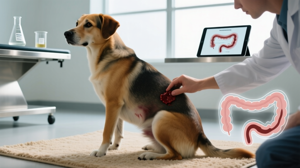
If you've ever glanced at your dog's stool and spotted bright red blood, your stomach probably dropped. Hematochezia — the veterinary term for fresh blood in feces — is one of the most common gastrointestinal complaints in veterinary medicine. According to data published in the Journal of Veterinary Internal Medicine, lower GI bleeding accounts for a significant percentage of canine emergency and urgent care visits each year. Understanding the physiological mechanism behind it, what causes it, and when it crosses from "monitor at home" to "drive to the vet now" is essential for every dog owner.
What Exactly Is Hematochezia? The Physiology of Lower GI Bleeding
Hematochezia (from Greek haima meaning blood and chēzia meaning to defecate) specifically refers to fresh, unmodified blood that appears bright red or maroon in the stool. This is a critical distinction from melena, where blood has traveled through the stomach and small intestine, becoming oxidized and appearing black and tarry.
The Anatomy of Canine Lower GI Bleeding
The canine gastrointestinal tract is roughly 3.5 times the body length — significantly shorter than the 6× body length in humans. This means transit time from ingestion to defecation averages 8 to 10 hours in dogs (compared to 24-72 hours in humans). When bleeding occurs in the descending colon, rectum, or anal region, blood has minimal contact time with digestive enzymes and bacteria, so it emerges looking essentially the same as when it entered the lumen.
The colon's mucosal lining is richly vascularized with a network of arterioles, capillaries, and venules just beneath a single layer of columnar epithelial cells. Any disruption to this thin barrier — whether from inflammation, ulceration, trauma, or parasite attachment — exposes these superficial blood vessels. Even minor mucosal damage can produce visible blood because the colonic vasculature is so close to the surface.
The canine colon also differs from the human colon in one key way relevant to hematochezia: dogs have a relatively shorter cecum and lack an appendix, but their ascending colon features a more prominent spiral fold pattern. This anatomical arrangement means that inflammatory or infectious processes in the canine colon tend to produce more diffuse mucosal irritation, leading to the characteristic "bloody mucus" presentation seen in conditions like hemorrhagic gastroenteritis (HGE), now more accurately termed acute hemorrhagic diarrhea syndrome (AHDS).
Why Do Dogs Develop Hematochezia? Common Causes Ranked by Frequency
Not all hematochezia is created equal. In a 2019 retrospective study published in the Journal of Small Animal Practice, researchers categorized the most common etiologies of canine hematochezia presented to referral hospitals. Here's what the data shows, organized from most to least common in general practice:
| Cause | Typical Presentation | Estimated Prevalence | Vet Visit Urgency |
|---|---|---|---|
| Colitis (inflammatory) | Mucus + blood streaks, frequent small stools, straining | ~30-35% of cases | Same-week appointment |
| Parasitic infection (hookworms, whipworms, Giardia) | Intermittent bloody diarrhea, weight loss, poor coat | ~20-25% of cases | Same-week appointment |
| Dietary indiscretion | Acute onset after eating unusual food, self-limiting | ~15-20% of cases | Monitor 24-48 hrs |
| AHDS (hemorrhagic gastroenteritis) | Sudden "raspberry jam" diarrhea, vomiting, dehydration | ~8-12% of cases | Emergency — same day |
| Anal gland disease | Blood on stool surface, scooting, foul odor | ~5-10% of cases | Routine appointment |
| Rectal polyps or neoplasia | Chronic intermittent bleeding, tenesmus (straining) | ~3-5% of cases | Prompt evaluation |
| Parvovirus | Profuse bloody diarrhea, vomiting, fever, lethargy in puppies | ~2-5% (unvaccinated pups) | Emergency — same day |
| Coagulopathy (rodenticide poisoning, clotting disorders) | Blood from multiple sites, bruising, weakness | ~1-3% of cases | Emergency — immediately |
The key takeaway: the pattern of bleeding matters more than the presence of bleeding. A single streak of bright red blood on a formed stool in an otherwise healthy adult dog is fundamentally different from a puppy producing liquid "raspberry jam" diarrhea.
The Evolutionary Context: Why Canine GI Tracts Are Vulnerable to Bleeding
Ancestral perspective: The domestic dog's gastrointestinal tract is essentially that of a facultative carnivore — an animal that evolved to eat primarily meat but can adapt to omnivorous diets when necessary. Gray wolves (Canis lupus), the primary ancestors of domestic dogs, consume prey whole: muscle meat, organs, bone, fur, and the semi-digested plant matter in the prey's gut.
This evolutionary diet shaped a GI tract with a highly acidic stomach (pH 1-2, compared to human pH 2-4), rapid transit time, and a relatively short colon designed to process protein and fat efficiently. The trade-off: the canine colonic mucosa is thinner and more reactive than that of true omnivores like humans or pigs. When modern dogs encounter dietary irritants, parasites, or pathogens that their ancestral diet didn't typically include in concentrated form — such as high-fiber commercial foods, chemical additives, or environmental contaminants — the colonic lining can mount an inflammatory response that manifests as hematochezia.
Additionally, ancestral canids practiced coprophagy (eating feces) and scavenged decomposing carcasses, meaning their immune systems evolved to handle high bacterial loads. Modern dogs retain this robust immune response, but when combined with less diverse gut microbiomes (a consequence of processed diets and antibiotic use), the colonic immune system can overreact to relatively minor irritants, triggering inflammatory colitis with secondary bleeding.
Research published in Nature Communications (2013, Axelsson et al.) demonstrated that domestic dogs evolved increased copy numbers of the amylase gene (AMY2B) compared to wolves, enabling better starch digestion. However, this adaptation occurred over only ~15,000-30,000 years of domestication — a blink of an eye in evolutionary terms. The colonic mucosa hasn't fully adapted to the high-carbohydrate, high-fiber diets found in many modern kibbles, which may partly explain the prevalence of dietary-related colitis and subsequent hematochezia in pet dogs.
How Is Canine Hematochezia Different From Humans?
While humans and dogs both experience lower GI bleeding, several physiological differences make hematochezia in dogs both more common and more diagnostically challenging:
| Factor | Dogs | Humans |
|---|---|---|
| GI transit time | 8-10 hours (fast) | 24-72 hours (slow) |
| Colonic mucosa thickness | Thinner, more reactive | Thicker, more resilient |
| Stomach pH | 1-2 (highly acidic) | 2-4 (moderately acidic) |
| Common causes | Parasites, dietary indiscretion, AHDS, parvovirus | Hemorrhoids, diverticulosis, IBD, colorectal cancer |
| Pain communication | Subtle behavioral changes, hiding, restlessness | Verbal reporting of cramping, urgency |
| Diagnostic approach | Fecal panel, bloodwork, imaging; colonoscopy less common | Colonoscopy is gold standard |
One significant difference: dogs cannot verbally report abdominal cramping, urgency, or the sensation of incomplete evacuation. This means owners must rely on behavioral cues — increased frequency of defecation, straining (tenesmus), restlessness, or sudden house-soiling in a previously house-trained dog — to recognize that something is wrong beyond the visible blood.
When Hematochezia Signals a Problem: Red Flags vs. Monitor-at-Home Scenarios
This is where most dog owners need the most guidance. Not every instance of blood in stool is an emergency, but some situations demand immediate action.
- Your dog is a puppy under 6 months with bloody diarrhea (parvovirus risk)
- Blood volume is large — more than a few streaks, or the stool is entirely liquid and bloody
- Stool resembles "raspberry jam" or dark grape jelly (AHDS indicator)
- Your dog is vomiting repeatedly alongside the bloody stool
- Gums appear pale, white, or gray (sign of significant blood loss)
- Your dog is lethargic, collapsed, or unresponsive
- You suspect rodenticide (rat poison) ingestion — call ASPCA Animal Poison Control: (888) 426-4435 (24/7, consultation fee applies) immediately
- Your dog is unvaccinated or overdue on DHPP vaccines
- A single episode of a small blood streak on an otherwise formed stool
- Your dog is an adult, fully vaccinated, and acting normally
- Appetite, energy level, and water intake are all normal
- No vomiting, no straining, and no repeated episodes within 24 hours
- You recently changed food brands (transitional GI upset is common)
What Your Veterinarian Will Do: Diagnostic Workup
When you bring your dog in for hematochezia, expect a systematic diagnostic approach. Here's what typically happens, along with realistic cost ranges (US averages, 2025-2026):
- Physical examination ($55-$95): Palpation of the abdomen, digital rectal exam, assessment of hydration status via skin tent test and gum moisture, and evaluation of mucous membrane color.
- Fecal flotation and direct smear ($35-$65): Checks for parasite eggs (hookworms, whipworms) and protozoal organisms (Giardia, Coccidia). A SNAP Giardia test ($40-$55) may be added.
- Complete blood count (CBC) and chemistry panel ($120-$220): Evaluates red blood cell count (to assess blood loss severity), white blood cell patterns (infection vs. inflammation), platelet count (clotting ability), and organ function.
- Canine parvovirus fecal antigen test ($40-$60): A rapid in-clinic test for puppies or unvaccinated dogs presenting with bloody diarrhea.
- Abdominal radiographs or ultrasound ($200-$500): Performed if a foreign body, intussusception (telescoping of the intestine), or mass is suspected.
- Colonoscopy with biopsy ($800-$1,500): Reserved for chronic, recurrent hematochezia that doesn't respond to empirical treatment. Usually performed at a referral center under general anesthesia.
Total cost for a standard workup (exam + fecal + bloodwork) typically runs $210-$380 at a general practice. Emergency clinic visits with IV fluid therapy for AHDS can range from $800-$2,500 depending on hospitalization duration.
Breed Variations: Which Dogs Are More Prone to Hematochezia?
While any dog can develop hematochezia, certain breeds show predispositions to conditions that commonly cause lower GI bleeding:
- Miniature Schnauzers, Yorkshire Terriers, and Toy Poodles: Higher incidence of AHDS (acute hemorrhagic diarrhea syndrome). These small breeds seem particularly susceptible to the rapid fluid shifts and colonic mucosal sloughing that characterize AHDS. A study in the Journal of Veterinary Emergency and Critical Care found that dogs under 20 lb (9 kg) were overrepresented in AHDS cases.
- German Shepherds and Boxers: Predisposed to inflammatory bowel disease (IBD), specifically lymphocytic-plasmacytic colitis, which causes chronic intermittent hematochezia. German Shepherds also have a documented breed-specific enteropathy with cobalamin malabsorption.
- French Bulldogs and other brachycephalic breeds: Higher rates of food sensitivities and chronic colitis, potentially linked to altered gut microbiome composition associated with brachycephalic anatomy and chronic low-grade hypoxia.
- Large and giant breeds (Great Danes, Irish Setters): More susceptible to rectal polyps and colorectal neoplasia in later life, which present as chronic hematochezia with tenesmus.
- Greyhounds and other sighthounds: Unique sensitivity to NSAIDs (non-steroidal anti-inflammatory drugs) that can cause GI ulceration and bleeding at doses tolerated by other breeds.
Common Misconceptions About Hematochezia in Dogs
| Myth | Fact |
|---|---|
| "A little blood in stool is always normal" | A single streak may be benign, but recurrent hematochezia — even small amounts — indicates ongoing mucosal damage that warrants investigation. Chronic colitis left untreated can lead to protein-losing enteropathy and weight loss. |
| "My dog ate something red, so it's just food coloring" | While red-pigmented foods (beets, red velvet cake) can temporarily tint stool, true hematochezia has a distinct appearance — blood coats or streaks the stool rather than uniformly coloring it. If you're unsure, place a sample on a white paper towel; blood will smear red, while food pigment typically creates an orange or pink stain. |
| "Hematochezia always means my dog has parvo" | Parvovirus is one cause, primarily in unvaccinated puppies under 6 months. In adult, vaccinated dogs, colitis, parasites, and dietary causes are far more common. Parvovirus also presents with a distinctive foul odor, severe lethargy, and rapid deterioration that differentiates it from milder causes. |
| "I can treat bloody stool with Pepto-Bismol" | Bismuth subsalicylate (Pepto-Bismol) contains salicylate, which can worsen GI bleeding and is toxic to dogs at incorrect doses. It also turns stool black, masking melena. Never give Pepto-Bismol for hematochezia. Consult your veterinarian before administering any over-the-counter medication. |
| "Fasting my dog for 24 hours will fix it" | While short-term fasting (12-24 hours) was historically recommended for GI upset, current veterinary consensus favors feeding a highly digestible, low-fat diet to maintain enterocyte nutrition. Prolonged fasting can actually impair intestinal mucosal healing. A bland diet of boiled chicken breast (no skin, no bones) and white rice in a 1:2 ratio is a better approach while awaiting your vet appointment. |
Practical Takeaways: What This Means for Your Daily Life
Prevention Strategies That Actually Work
- Maintain a consistent diet. Sudden food changes are a top trigger for dietary colitis. When transitioning foods, follow a 7-10 day gradual mix: 75% old/25% new for days 1-3, 50/50 for days 4-6, 25% old/75% new for days 7-9, then 100% new on day 10.
- Keep parasite prevention current. Monthly heartworm preventives like Heartgard Plus or Interceptor Plus also cover intestinal parasites (hookworms, roundworms, and in the case of Interceptor Plus, whipworms). Annual fecal exams catch infections before they cause clinical hematochezia.
- Supervise outdoor time. Dogs who scavenge garbage, eat wildlife feces, or drink from stagnant puddles have higher rates of parasitic and bacterial colitis. A reliable "leave it" cue trained with positive reinforcement is one of the best preventive tools you have.
- Consider a probiotic supplement. Products containing Enterococcus faecium (such as Purina FortiFlora) or multi-strain veterinary formulations (such as Proviable-DC) can support gut microbiome stability during diet transitions or stress. Evidence from the Merck Veterinary Manual supports probiotic use in managing acute colitis.
- Manage stress. Stress colitis is well-documented in dogs. Boarding, travel, new household members, and loud events (fireworks, thunderstorms) can trigger inflammatory responses in the colonic mucosa. If your dog is stress-prone, discuss situational anti-anxiety options with your veterinarian before known triggers.
What to Do When You Spot Blood
- Take a photo of the stool in natural light. Your vet will find this extremely helpful — color, volume, and consistency are hard to describe accurately from memory.
- Collect a fresh sample (less than 4 hours old) in a clean plastic bag or container. Keep it cool (refrigerate, don't freeze) and bring it to your appointment.
- Note the context: What did your dog eat in the last 24 hours? Any new treats, table scraps, or found items? Any recent medication changes (especially NSAIDs like carprofen or meloxicam)?
- Monitor frequency: Count how many bowel movements occur in the next 12 hours and whether each contains blood. This timeline helps your vet differentiate acute from chronic causes.
- Do NOT administer human medications — no Pepto-Bismol, no Imodium (loperamide can be dangerous in certain breeds with the MDR1 gene mutation, including Collies, Australian Shepherds, and related herding breeds), and no aspirin.
Related Conditions and Cross-References
Understanding hematochezia benefits from knowing related GI conditions that dog owners should be aware of:
- Melena: Dark, tarry stool indicating upper GI bleeding (stomach or small intestine). Often caused by NSAID-induced ulcers, foreign bodies, or clotting disorders. This is generally more serious than hematochezia because the bleeding source is higher in the GI tract.
- Acute Hemorrhagic Diarrhea Syndrome (AHDS): A life-threatening condition causing sudden, profuse "raspberry jam" diarrhea with rapid dehydration. PCV (packed cell volume) often exceeds 60% due to hemoconcentration. Requires aggressive IV fluid therapy; mortality is low (~5%) with prompt treatment but can be fatal without it.
- Hematochezia vs. rectal bleeding from trauma: If your dog recently had a rectal thermometer insertion, anal gland expression, or ingested a sharp object (bone fragments, sticks), the bleeding source may be mechanical rather than inflammatory. The treatment approach differs significantly.
- Chronic colitis: When hematochezia recurs over weeks or months, your vet may investigate food-responsive enteropathy (trial an elimination diet with a novel protein like rabbit, venison, or hydrolyzed protein), antibiotic-responsive diarrhea (tylosin or metronidazole trial), or immune-mediated IBD requiring immunosuppressive therapy.
Frequently Asked Questions
Is hematochezia in dogs always an emergency?
No. A single, small streak of bright red blood on an otherwise normal stool in a healthy, vaccinated adult dog who is eating and acting normally is usually not an emergency. Monitor for 24-48 hours. If it recurs, increases in volume, or is accompanied by other symptoms (vomiting, lethargy, diarrhea, loss of appetite), contact your veterinarian promptly.
Can stress cause bloody stool in dogs?
Yes. Stress colitis is a well-documented phenomenon in dogs. The mechanism involves the brain-gut axis: psychological stress triggers cortisol and catecholamine release, which alters colonic motility, increases mucosal permeability, and disrupts the gut microbiome. Boarding, moving, new pets, loud noises, and separation anxiety are common triggers. Stress-related hematochezia typically resolves once the stressor is removed, but chronic stress can lead to persistent colitis requiring veterinary management.
What does it mean when my dog's stool is coated in mucus and blood?
Mucus production is the colon's protective response to inflammation. When the colonic mucosa is irritated — whether by parasites, bacteria, dietary irritants, or immune-mediated processes — goblet cells increase mucus secretion to coat and protect the damaged surface. Blood mixed with mucus (often described as "bloody jelly") is a hallmark of colitis and warrants a veterinary visit for fecal testing and appropriate treatment.
How is hematochezia diagnosed differently from melena?
Your veterinarian distinguishes them visually first: hematochezia is bright red (fresh blood, lower GI source), while melena is black and tarry (digested blood, upper GI source). Diagnostic workups differ accordingly — hematochezia prompts fecal analysis, rectal examination, and possibly colonoscopy, while melena prompts upper GI investigation including gastric ultrasound, coagulation testing, and potentially endoscopy of the stomach and duodenum.
Can my dog get hematochezia from eating bones?
Yes. Cooked bones (especially chicken, pork, and rib bones) can splinter into sharp fragments that mechanically lacerate the colonic and rectal mucosa during passage. This produces hematochezia that may appear 12-48 hours after ingestion. Raw bones are less likely to splinter but still carry risk. If your dog has eaten cooked bones and develops bloody stool, contact your veterinarian — radiographs may be needed to check for obstruction or perforation.
What is the prognosis for dogs with hematochezia?
Prognosis depends entirely on the underlying cause. Dietary indiscretion and mild colitis typically resolve within 3-5 days with dietary management. Parasitic infections clear with appropriate deworming (e.g., fenbendazole at 50 mg/kg daily for 3-5 days). AHDS has a good prognosis (~95% survival) with prompt aggressive fluid therapy. Parvovirus survival rates are approximately 80-95% with intensive care but drop to below 50% without treatment. Colorectal tumors carry a variable prognosis depending on staging and surgical resectability. Your veterinarian will discuss expected outcomes once a diagnosis is established.
References
Robin Maitland
All our authors care for dogs every day — read more of their work on the authors page.


