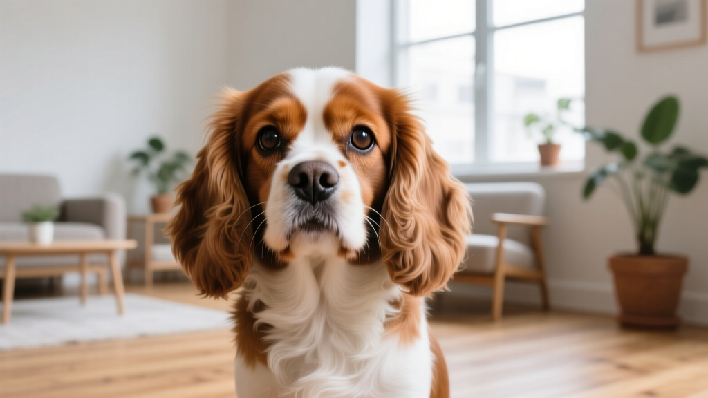
Eye Snot in Dogs: The Science of Canine Eye Discharge Explained
Why do dogs get eye snot? Learn the canine science behind eye discharge, when it's normal, breed risks, and how to tell healthy tears from infection.

What Exactly Is Eye Snot in Dogs?
That gunky buildup you wipe from your dog's eyes every morning isn't random — it's the byproduct of a sophisticated ocular maintenance system working around the clock. The canine eye produces three distinct tear layers, each serving a specific function, and "eye snot" is what's left over when those layers do their job.
The Three-Layer Tear Film
The canine tear film consists of:
- Lipid layer (outermost): Produced by the meibomian glands along the eyelid margins. This oily coating prevents tear evaporation and keeps the eye surface lubricated.
- Aqueous layer (middle): Secreted by the lacrimal gland and the gland of the third eyelid (nictitans gland). This watery layer delivers oxygen and nutrients to the cornea and flushes away debris.
- Mucin layer (innermost): Produced by conjunctival goblet cells. Mucin anchors the tear film to the hydrophobic corneal surface, ensuring even coverage.
When your dog sleeps, blinking stops. Without the mechanical action of eyelids spreading fresh tears and draining old ones through the nasolacrimal ducts, the aqueous layer evaporates while mucin, lipids, exfoliated epithelial cells, and trapped environmental dust concentrate in the medial canthus (inner eye corner). That concentrated residue is what owners colloquially call "eye snot."
The Merck Veterinary Manual describes this tear film system as essential to corneal health — without adequate production of all three layers, dogs develop keratoconjunctivitis sicca (dry eye), which paradoxically produces more thick, ropey discharge because the eye compensates with excess mucus when aqueous tears are insufficient.
Why Do Dogs Produce Eye Discharge? The Evolutionary Context
In wild canids — gray wolves, coyotes, foxes — the eye sits in a properly proportioned orbit with a functional nasolacrimal punctum (the tiny drainage hole on the inner eyelid). Tears drain efficiently from the eye surface through the puncta, into the nasolacrimal duct, and out through the nose. This is why you'll rarely see wild canids with visible eye discharge in field photography.
Domestication changed this equation. Approximately 15,000–30,000 years of selective breeding produced skull shapes that wolves never possessed. Brachycephalic breeds (Pugs, Bulldogs, Shih Tzus, Pekingese) have shallow orbits, prominent globes, and compressed nasolacrimal ducts that often don't drain properly. The result: tears overflow onto the face (epiphora), creating a moist environment where porphyrin pigments in the tears oxidize and stain the fur reddish-brown, and discharge accumulates far more than in mesocephalic or dolichocephalic breeds.
How Is Canine Eye Discharge Different from Humans?
Both humans and dogs produce rheum (the technical term for sleep crust), but there are key physiological differences:
| Feature | Dogs | Humans |
|---|---|---|
| Third eyelid | Present — nictitating membrane actively sweeps debris and contributes ~30-50% of tear production via the nictitans gland | Vestigial (plica semilunaris) — non-functional, produces negligible tears |
| Tear composition | Higher mucin concentration; more prone to ropey discharge when aqueous layer is deficient | More balanced aqueous-to-mucin ratio in healthy individuals |
| Porphyrin staining | Significant — tears contain porphyrins that oxidize to reddish-brown on light fur | Minimal — human tears contain far lower porphyrin concentrations |
| Nasolacrimal anatomy | Single punctum per eyelid in most breeds; brachycephalic breeds often have congenital punctal aplasia (missing drainage holes) | Two puncta (upper and lower); congenital absence is rare |
| Blink rate | Approximately 10-15 blinks per minute (varies by breed and activity) | Approximately 15-20 blinks per minute |
The third eyelid distinction matters practically: if your dog's third eyelid becomes visibly protruded (sometimes called "cherry eye" when the gland prolapses, or simply elevated when the dog is unwell), it can disrupt normal tear distribution and increase discharge accumulation.
Breed Variations: Which Dogs Are Prone to Excessive Eye Discharge?
Not all eye snot is created equal. Breed conformation plays the single largest role in how much discharge your dog produces and whether it signals a problem.
High-Discharge Breeds (Structural Predisposition)
- Brachycephalic breeds — Pugs, English Bulldogs, French Bulldogs, Shih Tzus, Pekingese, Boston Terriers: Shallow orbits cause prominent eyes (exophthalmos), incomplete eyelid closure during sleep (lagophthalmos), and kinked or absent nasolacrimal ducts. Expect daily discharge as a baseline.
- Breeds with excessive facial skin folds — Bloodhounds, Basset Hounds, Cocker Spaniels, Shar-Peis: Skin folds trap moisture and debris near the eyes, creating a warm environment for bacterial overgrowth. Entropion (inward-rolling eyelids) is also common, causing lashes to rub the cornea and stimulate excess mucus production.
- Breeds predisposed to keratoconjunctivitis sicca (KCS/dry eye) — Cavalier King Charles Spaniels, English Bulldogs, West Highland White Terriers, American Cocker Spaniels: Immune-mediated destruction of lacrimal tissue reduces aqueous tear production. The eye compensates with thick, yellow-green mucoid discharge. KCS affects an estimated 1-4% of the general dog population but prevalence in predisposed breeds may reach 10-20%.
Low-Discharge Breeds
- Dolichocephalic breeds — Greyhounds, Borzois, Salukis, Whippets: Long muzzles and properly proportioned orbits mean tears drain efficiently. Visible discharge beyond a tiny morning crust warrants investigation.
- Mesocephalic working breeds — Labrador Retrievers, German Shepherds, Border Collies: Moderate skull proportions generally support normal tear drainage, though individual variation exists.
When Eye Snot Signals a Problem: A Diagnostic Framework
The color, consistency, and volume of your dog's eye discharge tell a detailed story about what's happening beneath the surface. Here's how veterinary ophthalmologists interpret these signals:
| Discharge Type | Appearance | Likely Causes | Urgency |
|---|---|---|---|
| Clear/watery | Thin, transparent fluid | Allergies, irritants (dust, wind), early viral conjunctivitis, blocked tear duct overflow | Monitor 24-48 hours; see vet if persistent |
| White/gray mucoid | Stringy or ropey, white to light gray | Dry eye (KCS), mild conjunctivitis, normal sleep accumulation in small amounts | Schedule vet visit within 1 week if new or increasing |
| Yellow/green purulent | Thick, opaque, yellow-green; may crust heavily | Bacterial conjunctivitis, corneal ulcer with secondary infection, advanced KCS, dacryocystitis (tear duct infection) | See vet within 24-48 hours |
| Bloody/brown-tinged | Reddish-brown or blood-flecked discharge | Trauma, corneal ulcer, foreign body, neoplasia (tumor) | Emergency — see vet immediately |
| Reddish-brown staining | Not discharge per se — oxidized porphyrin staining on fur below the eye | Chronic epiphora (tear overflow), common in light-colored brachycephalic breeds and small breeds like Maltese and Bichon Frisés | Cosmetic concern; address underlying overflow with vet |
- Your dog is squinting, holding the eye shut, or pawing at the eye (signs of corneal pain)
- The eye appears cloudy, blue, or has a visible dent/irregularity on the corneal surface
- Discharge is accompanied by lethargy, fever, loss of appetite, or nasal discharge (possible systemic infection such as canine distemper)
- You suspect a foreign body (foxtail, grass seed, plant material) is lodged under the eyelid
- The eye is visibly bulging or the third eyelid is covering more than 50% of the globe
Corneal ulcers can progress to perforation within 24-72 hours without treatment. Never wait to "see if it gets better" when pain signs are present.
Common Misconceptions About Dog Eye Discharge
| Myth | Fact |
|---|---|
| "Eye boogers mean my dog is sick" | Small amounts of clear-to-gray discharge after sleep are a normal byproduct of tear film turnover. Just as humans wake with sleep crust, dogs accumulate rheum. It only indicates illness when volume, color, or consistency changes significantly. |
| "I should use human eye drops on my dog" | Human over-the-counter eye drops like Visine (tetrahydrozoline) constrict blood vessels and are not formulated for canine ocular pH or tear film composition. Some contain preservatives that can damage the canine cornea. Only use veterinary-prescribed ophthalmic medications. Sterile saline eyewash (without active drugs) is generally safe for flushing debris, but consult your veterinarian before applying anything medicated. |
| "Reddish-brown tear stains mean my dog has a yeast infection" | Tear staining is primarily caused by porphyrins — iron-containing molecules excreted in tears, saliva, and urine. When tears sit on light-colored fur and are exposed to air, porphyrins oxidize to a rust-brown color. While secondary yeast (Malassezia) overgrowth can occur in the moist environment, the staining itself is a chemical reaction, not an infection. Addressing the overflow (epiphora) is the primary solution. |
| "Cleaning my dog's eyes daily will cause irritation" | Gentle daily cleaning with a soft, lint-free cloth moistened with sterile saline or a veterinary ophthalmic wipe (e.g., Vetericyn Eye Wash, Nutri-Vet Eye Rinse) is beneficial, especially for brachycephalic breeds. The key is technique: wipe from the inner corner outward, use a fresh section of cloth for each eye, and avoid touching the corneal surface directly. |
| "All eye discharge is conjunctivitis" | Conjunctivitis (inflammation of the conjunctiva) is one cause, but discharge also results from KCS, corneal ulcers, uveitis, glaucoma, nasolacrimal duct obstruction, entropion, ectopic cilia, foreign bodies, and systemic diseases like canine distemper or leishmaniasis. Accurate diagnosis requires veterinary examination, often including a Schirmer tear test, fluorescein stain, and tonometry (intraocular pressure measurement). |
Practical Eye Care: What This Means for Your Daily Routine
Daily Eye Maintenance Protocol
For most dogs (mesocephalic/dolichocephalic, no pre-existing conditions):
- Check eyes once daily — ideally during a morning grooming or feeding routine.
- If you see a small crust at the inner canthus, moisten a clean gauze pad or lint-free cloth with sterile saline (available at pharmacies for ~$3-5 per bottle).
- Hold the pad against the crust for 5-10 seconds to soften it, then gently wipe from the inner corner toward the outer corner in a single stroke.
- Use a fresh pad for the other eye to avoid cross-contamination.
- No daily cleaning is needed if eyes are clear and discharge-free.
For brachycephalic and high-discharge breeds:
- Clean eyes twice daily (morning and evening).
- Trim hair around the eyes weekly to prevent wicking of tears onto the face. Use blunt-nosed grooming scissors — never sharp-tipped scissors near the eye.
- Apply a veterinary-approved eye lubricant (e.g., Puralube Vet Ointment, ~$12-18 per tube) at bedtime if your vet has recommended it for lagophthalmos (incomplete eyelid closure).
- Inspect facial folds below the eyes daily; clean and dry them to prevent fold dermatitis, which can secondarily affect the eye.
- Schedule veterinary ophthalmic checkups every 6-12 months, including a Schirmer tear test (~$25-50 per eye at most general practices) to catch KCS early.
What to Expect at the Vet
If you bring your dog in for abnormal eye discharge, here's the typical diagnostic sequence and approximate costs (US averages, 2025-2026):
- Ophthalmic exam: $55-90 (general practice) or $150-250 (veterinary ophthalmologist specialist)
- Schirmer tear test (STT): Measures aqueous tear production in mm/min. Normal is >15 mm/min; values below 10 mm/min indicate KCS. Cost: $25-50.
- Fluorescein stain: Orange dye applied to the cornea that highlights ulcers or abrasions under blue light. Cost: $20-40.
- Tonometry: Measures intraocular pressure to rule out glaucoma (normal canine IOP: 10-25 mmHg). Cost: $30-60.
- Nasolacrimal flush: Saline flushed through the tear ducts to check for obstruction. Cost: $40-80.
- Cytology/culture: If purulent discharge is present, a swab may be cultured to identify the bacterial species and determine antibiotic sensitivity. Cost: $50-120.
Total first-visit cost for a discharge workup typically ranges from $150-400 at a general practice. Treatment costs vary widely: topical antibiotic drops (e.g., neomycin-polymyxin-bacitracin ophthalmic ointment, ~$15-30 per tube) for bacterial conjunctivitis versus lifelong cyclosporine or tacrolimus therapy ($30-80/month) for KCS.
Related Ocular Behaviors and Abilities
Understanding eye discharge connects to several other aspects of canine eye function and behavior:
- Squinting (blepharospasm): This is a pain response, not a "sleepy" behavior. Dogs squint when the cornea is irritated, ulcerated, or when intraocular pressure is elevated. Any squinting lasting more than a few hours warrants veterinary examination.
- Third eyelid elevation: The nictitating membrane can passively elevate when the eye sinks into the orbit (due to dehydration, weight loss, or pain) or when the sympathetic nervous system is disrupted (Horner's syndrome). It can also actively sweep across the eye in response to irritation.
- Tear production and emotion: A 2022 study published in Current Biology found that dogs produce more tears during reunions with their owners compared to reunions with familiar non-owner humans, suggesting a link between oxytocin release and lacrimal secretion. However, dogs do not "cry" emotional tears in the way humans do — visible tear overflow is almost always a physical, not emotional, phenomenon.
- Canine vision and discharge impact: Heavy discharge crusted over the cornea physically obstructs light entry, temporarily reducing visual acuity. For working dogs (herding, detection, service work), even mild chronic discharge can impair performance. Regular cleaning is functionally important, not just cosmetic.
Frequently Asked Questions
Is eye snot in dogs normal?
Yes, in small quantities. A small amount of clear to light gray discharge in the inner corner of the eye after sleeping is a normal accumulation of tear film debris. It becomes abnormal when the volume increases noticeably, the color shifts to yellow or green, the consistency becomes thick and ropey, or your dog shows signs of discomfort (squinting, rubbing, redness).
Can dogs get pink eye from eye discharge?
Conjunctivitis (pink eye) causes discharge rather than the reverse. However, chronic untreated discharge creates a moist environment where bacteria can proliferate, potentially leading to secondary conjunctivitis or skin infections (moist dermatitis) on the face below the eye. Canine conjunctivitis is rarely contagious to humans, but bacterial conjunctivitis can spread between dogs in close contact.
How do dogs process tear drainage differently than humans?
Dogs have a single lacrimal punctum on each eyelid (upper and lower), though in brachycephalic breeds the upper punctum is frequently absent (punctal aplasia). Tears drain through the puncta into the nasolacrimal duct, which empties into the nasal cavity — the same endpoint as in humans, but the canine duct takes a more tortuous path through the maxillary bone. This anatomical complexity means obstructions are more common in dogs, especially in breeds with compressed facial skeletons.
What does it mean when my dog has one eye with more discharge than the other?
Unilateral (one-sided) discharge often points to a localized problem: a foreign body trapped under that eyelid, a corneal scratch on that eye, a blocked nasolacrimal duct on that side, or an entropion affecting only one eyelid. Bilateral (both eyes) discharge more commonly suggests systemic causes like allergies, KCS, or infectious conjunctivitis. Either way, asymmetric discharge warrants a veterinary exam to identify the specific cause.
Can I use coconut oil or apple cider vinegar to clean my dog's eyes?
No. Neither coconut oil nor apple cider vinegar is appropriate for ocular use. Coconut oil is not sterile and can introduce bacteria to the eye surface; its fatty composition also disrupts the lipid layer of the tear film. Apple cider vinegar is acidic (pH 2-3) and will cause significant corneal irritation and potential chemical burns. Stick to sterile saline or veterinary-formulated ophthalmic rinses that are pH-balanced for the canine eye (approximately pH 7.4-7.8).
Key Takeaways
- Eye snot is a normal byproduct of the canine tear film system — small amounts of clear/gray discharge after sleep are not a concern.
- Breed conformation is the biggest factor in discharge volume: brachycephalic breeds require daily eye maintenance as a lifelong commitment.
- Color changes (yellow, green, bloody) and behavioral signs (squinting, pawing) are your primary indicators that veterinary attention is needed.
- Never use human eye drops, home remedies, or unverified products in your dog's eyes — the cornea is delicate and easily damaged.
- Budget $150-400 for a diagnostic workup if discharge is abnormal, and factor in ongoing costs for chronic conditions like KCS ($30-80/month for medication).
- Consult your veterinarian before starting any eye care regimen beyond simple saline wiping — early detection of KCS, glaucoma, or corneal disease can save your dog's vision.
Robin Maitland
All our authors care for dogs every day — read more of their work on the authors page.