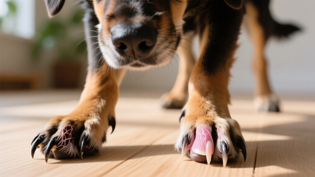
How Canine Limbs Are Built for Speed and Agility
Explore dog leg anatomy in depth — bones, joints, muscles, and evolutionary design. Learn how canine limbs work and what it means for your dog's health.

The Quick Answer: How Is Dog Leg Anatomy Structured?
Dogs are digitigrade animals, meaning they walk on their toes rather than the soles of their feet like humans. Each front leg contains the equivalent of a human arm — humerus, radius, ulna, carpal bones, metacarpals, and phalanges — while each rear leg mirrors the human leg with a femur, tibia, fibula, tarsal bones, metatarsals, and phalanges. The canine hindlimb generates the majority of propulsive force during running, while the forelimbs absorb impact and provide steering. Dogs have a vestigial clavicle (collarbone) that is not attached to the skeleton, allowing greater stride length. Most dogs have 18 toes total (five on each front paw including the dewclaw, and four on each rear paw), though some breeds possess additional rear dewclaws.
Understanding dog leg anatomy isn't just academic curiosity — it directly affects how you exercise your dog, recognize injury, choose the right activities for your breed, and advocate for your dog at the veterinary clinic. Whether you're watching a Greyhound explode off the starting line or a Dachshund navigate stairs, you're witnessing millions of years of evolutionary engineering at work.
The Skeletal Framework: Bones of the Canine Leg
The canine appendicular skeleton — the limbs and their attachment points — contains roughly 80 of the dog's total 319–321 bones (count varies slightly by breed and dewclaw presence). Understanding the bone structure from shoulder to paw gives you a mental map for identifying where injuries occur.
Forelimb Bones (Front Legs)
| Bone | Human Equivalent | Function & Clinical Notes |
|---|---|---|
| Scapula | Shoulder blade | Floats on muscle — no true bony joint to the ribcage. Allows tremendous forelimb reach. |
| Humerus | Upper arm | Single long bone. Fractures uncommon in adults but seen in toy-breed puppies. |
| Radius & Ulna | Forearm | Two parallel bones. The ulna forms the elbow point (olecranon). Common fracture site in jumping dogs. |
| Carpal Bones (6–7) | Wrist | Arranged in two rows. Hyperextension injuries occur in agility dogs landing incorrectly. |
| Metacarpals (5) | Palm bones | Weight-bearing in digitigrade stance. Metacarpals 3 and 4 carry the most load. |
| Phalanges (14+) | Finger bones | Three per digit (proximal, middle, distal). The distal phalanx supports the claw. |
Hindlimb Bones (Rear Legs)
The rear limb connects to the spine via the pelvis (os coxae), a fused structure of ilium, ischium, and pubis. This solid connection to the axial skeleton is why hindlimbs generate so much more driving force than forelimbs.
- Femur — The longest bone in the dog's body. The femoral head articulates with the acetabulum of the pelvis to form the hip joint. This is the primary site affected by hip dysplasia.
- Patella — A sesamoid bone embedded in the quadriceps tendon. Luxating patellas (kneecaps that pop out of place) are extremely common in small breeds — the Orthopedic Foundation for Animals reports prevalence exceeding 7% in Pomeranians and Yorkshire Terriers.
- Tibia & Fibula — The shin bones. The tibial plateau angle is critical in cranial cruciate ligament (CCL) disease — dogs with steeper angles face higher rupture risk.
- Tarsal Bones (7) — The hock (ankle) complex. The calcaneus (heel bone) provides the lever arm for the Achilles tendon.
- Metatarsals & Phalanges — Structurally identical to the front paw but typically bearing four digits instead of five.
🐺 Evolutionary Context: Why Dogs Walk on Their Toes
Digitigrade locomotion evolved in cursorial (running-adapted) predators. By standing on their toes, ancestral canids effectively added an extra limb segment to their stride — the metacarpals and metatarsals function like extensions of the leg rather than a flat foot. This design increases stride length by approximately 20–30% compared to a plantigrade (flat-footed) stance. The tradeoff is reduced stability at rest, which is why dogs don't stand as effortlessly as humans. The vestigial clavicle — a small bony fragment embedded in the brachiocephalicus muscle — further extends reach by freeing the scapula from rigid skeletal attachment. This same evolutionary pressure shaped the Greyhound's extreme angulation, where shoulder and hip angles maximize the suspension phase of the gallop.
Joint Mechanics and the Stifle: Where Problems Concentrate
The canine stifle (knee) is the most complex and injury-prone joint in the dog's body. It is the anatomical equivalent of the human knee but operates at a fundamentally different angle — dogs stand with their stifles flexed, placing constant tension on the cranial cruciate ligament (CCL), the equivalent of the human ACL.
The Cranial Cruciate Ligament Problem
Research published in the Journal of the American Veterinary Medical Association estimates that cranial cruciate ligament disease affects approximately 2–5% of all dogs and is the single most common orthopedic surgery performed in veterinary medicine. Unlike human ACL tears, which often result from a single traumatic event, canine CCL disease is typically degenerative — the ligament slowly weakens over months or years before partial or complete rupture.
Key risk factors identified in veterinary literature:
- Body weight: Dogs over 50 lb (23 kg) face significantly elevated risk
- Tibial plateau angle: Angles exceeding 25° increase shear force on the CCL
- Breed predisposition: Labrador Retrievers, Rottweilers, and Newfoundlands show 2–5× higher incidence
- Neuter status: Early spay/neuter (before skeletal maturity) may increase risk in large breeds, per UC Davis research on Golden Retrievers and German Shepherds
- Obesity: Every additional pound of body weight multiplies joint loading by 4–6× during running
Other Critical Joints
| Joint | Type | Common Issues |
|---|---|---|
| Shoulder (glenohumeral) | Ball-and-socket | OCD (osteochondritis dissecans) in large-breed puppies; bicipital tenosynovitis in agility dogs |
| Elbow | Hinge (complex) | Elbow dysplasia — fragmented coronoid process, ununited anconeal process. Highly heritable in Retrievers and German Shepherds |
| Hip (coxofemoral) | Ball-and-socket | Hip dysplasia, Legg-Calvé-Perthes disease in small breeds, luxation from trauma |
| Hock (tarsocrural) | Hinge | Achilles tendon rupture in athletic dogs; tarsal hyperextension from falls |
Muscle Architecture and Locomotion: How Dogs Generate Movement
Canine locomotion depends on a proximal-distal muscle distribution — the largest, most powerful muscles cluster near the body (gluteals, quadriceps, hamstrings in the rear; pectorals, deltoids, and triceps in the front), while the distal limb (below the knee and elbow) is mostly tendon and bone. This design minimizes the weight the dog must swing with each stride, reducing metabolic cost during sustained running.
How Dogs Move: Gait Breakdown
Dogs use four primary gaits, each engaging the leg anatomy differently:
- Walk (3–4 mph / 5–6 km/h) — A four-beat gait with three feet on the ground at all times. Minimal joint flexion; mostly used for patrolling and scent investigation.
- Trot (5–8 mph / 8–13 km/h) — A two-beat diagonal gait (left front + right hind move together). This is the most energy-efficient gait for medium distances and the gait judges evaluate in conformation shows. The Merck Veterinary Manual notes that efficient trotting breeds like German Shepherds have evolved angulation (joint angles) that maximizes stride length at this gait.
- Gallop (15–45 mph / 24–72 km/h depending on breed) — A four-beat gait with a full suspension phase where all four feet leave the ground. Greyhounds reach 45 mph (72 km/h) using a double suspension rotary gallop — two moments per stride where the dog is fully airborne.
- Canter (a transitional gait) — A three-beat asymmetric gait sometimes used between trot and gallop, particularly in herding dogs adjusting speed.
Breed Variations: How Leg Anatomy Differs Across Dogs
Selective breeding has dramatically modified canine leg proportions, sometimes in ways that create health vulnerabilities. The term chondrodysplastic refers to breeds with genetically shortened legs — a mutation in the FGF4 gene that causes abnormal cartilage development.
| Conformation Type | Example Breeds | Leg Anatomy Traits | Health Implications |
|---|---|---|---|
| Sighthound (dolichomorphic) | Greyhound, Whippet, Saluki | Extreme rear angulation, long metatarsals, deep chest, minimal body fat over joints | Higher CCL rupture speed; thin tissue prone to abrasions; anesthesia sensitivity |
| Chondrodysplastic (dwarf) | Dachshund, Corgi, Basset Hound | Shortened radius/ulna and tibia/fibula; normal-length spine; bowing of forelimbs | IVDD risk 10–12× higher than non-chondrodysplastic breeds; elbow incongruity; premature arthritis |
| Giant Breed | Great Dane, Mastiff, Saint Bernard | Massive long bones, slower skeletal maturation (18–24 months to full growth plate closure) | Osteosarcoma risk; panosteitis during growth; CCL disease; hip/elbow dysplasia |
| Working/Athletic | Border Collie, Belgian Malinois, Labrador Retriever | Moderate angulation, balanced proportions, strong musculature | Generally lower orthopedic disease rates; soft tissue injuries from high-intensity activity |
Practical Takeaways: What Leg Anatomy Means for Your Dog's Daily Life
Actionable Guidance Based on Canine Leg Structure
1. Respect growth plate closure timelines. Small breeds close growth plates around 8–10 months. Medium breeds: 12–14 months. Large/giant breeds: 18–24 months. Forced repetitive exercise (long-distance running, agility jumping) before closure can cause microfractures and growth deformities. For a 6-month-old Labrador, limit structured exercise to roughly 25–30 minutes of free-play walking per day — the "5 minutes per month of age" rule is a reasonable starting guideline.
2. Manage body weight ruthlessly. A dog at ideal body condition score (BCS 4–5/9) has roughly 40% less orthopedic disease risk than an overweight dog (BCS 7+/9). For a 65 lb (29 kg) Labrador, even 5 extra pounds means 20–30 lb of additional force on the stifles during running. Use a measuring cup for kibble — eyeballing portions leads to 20–30% overfeeding in most households.
3. Choose surfaces wisely. Concrete and asphalt provide zero shock absorption. For dogs with existing joint disease or breeds predisposed to CCL issues, prioritize grass, dirt trails, or rubber-matted surfaces. If you must walk on pavement, consider paw protection boots with cushioned soles (e.g., Ruffwear Grip Trex, approximately $90–$100/pair).
4. Warm up before intense activity. Just as human athletes warm up, dogs benefit from 5–10 minutes of walking before fetch sessions, agility runs, or dock diving. Cold muscles and tendons are more injury-prone — canine muscle reaches optimal working temperature around 101–102°F (38.3–38.9°C), slightly above resting.
5. Learn normal gait patterns. Watch your dog trot toward you on a smooth surface. The front and rear feet should track in a straight line (or slightly converging at speed). Any sudden head-bob, shortened stride, or "bunny-hopping" in the rear warrants a veterinary orthopedic exam within 48 hours.
Common Misconceptions About Dog Leg Anatomy
| Myth | Fact |
|---|---|
| "Dogs have four knees" | Dogs have two knees (stifles) on the rear legs and two elbows on the front legs. The hock (ankle) is often mistaken for a rear knee. |
| "Dewclaws are useless and should always be removed" | Front dewclaws attach via bone and tendon and serve a function — stabilizing the carpus during high-speed turning. The AVMA notes cosmetic dewclaw removal is controversial. Rear dewclaws (often loosely attached by skin only) are more prone to injury and may warrant removal. |
| "A limping dog is always in pain" | Limping can result from pain, but also from mechanical issues (luxating patella that isn't painful), neurological deficits (degenerative myelopathy), or even learned behavior. Always have persistent lameness (>48 hours) evaluated. |
| "Dogs' legs work just like human arms and legs" | While the bones are homologous (same evolutionary origin), the digitigrade stance, absent functional clavicle, and different joint angles make canine biomechanics fundamentally distinct. Comparing a dog's hock to a human knee is anatomically incorrect — the hock corresponds to the human ankle. |
| "If my dog is running fine, their legs are healthy" | Dogs mask pain exceptionally well — an evolutionary holdover from pack survival. Many dogs with partial CCL tears or early hip dysplasia will continue to run but show subtle gait changes visible only on veterinary exam or gait analysis. |
When Leg Anatomy Signals a Problem: Red Flags for Owners
⚠️ When to Call Your Veterinarian
Consult your veterinarian promptly if you observe any of the following. These signs may indicate orthopedic, neurological, or systemic disease requiring professional diagnosis — never attempt to diagnose or treat leg issues at home without veterinary guidance.
- Non-weight-bearing lameness (leg held completely off the ground) lasting more than 24 hours
- Sudden yelping during activity followed by refusal to use a limb
- Visible swelling of any joint, especially if warm to the touch
- "Bunny-hopping" gait in the rear — both hind legs moving together instead of alternating (common sign of hip dysplasia or bilateral CCL disease)
- Dragging toes or knuckling (walking on the top of the paw) — suggests neurological involvement such as IVDD or degenerative myelopathy
- Reluctance to jump into the car, climb stairs, or rise from rest — especially in dogs over 7 years old
- Asymmetric muscle loss in one hind leg compared to the other (visible thigh thinning) — indicates chronic disuse from pain
Emergency situations requiring same-day evaluation: inability to stand, sudden paralysis of rear legs, limb held at an abnormal angle (possible fracture), or a joint that appears to be dislocated.
Frequently Asked Questions About Dog Leg Anatomy
Why do dogs have a "backward knee" on their hind legs?
That apparent backward knee is actually the hock (tarsocrural joint), which corresponds to the human ankle, not the knee. The dog's true knee (stifle) is the forward-bending joint higher up on the hind leg, often hidden by fur and musculature. The hock bends backward because dogs stand on their toes with the ankle elevated — the same joint that bends forward in humans when you stand on your tiptoes.
Can dogs dislocate their legs?
Yes. The most common joint luxations in dogs are patellar luxation (kneecap slipping out of its groove — graded I through IV by severity) and hip luxation (femoral head popping out of the acetabulum, usually from trauma like being hit by a car). Patellar luxation is often congenital and bilateral in small breeds. Grades I–II may be managed conservatively; grades III–IV typically require surgical correction (trochleoplasty or tibial tuberosity transposition), with costs ranging from $1,500–$3,500 per knee depending on the procedure and region.
How do dogs process the mechanics of running so fast?
Canine locomotion is controlled by central pattern generators (CPGs) — neural circuits in the spinal cord that produce rhythmic stepping patterns without requiring conscious thought from the brain. This is why dogs can run, trot, and gallop fluidly while focused on a scent or prey. The cerebellum fine-tunes coordination and balance, while proprioceptors in muscles, tendons, and joints provide constant feedback about limb position. This decentralized control system allows reaction times far faster than conscious processing would permit.
What does it mean when my dog kicks their back legs after pooping?
This ground-scratching behavior isn't about covering waste — it's a visual and scent-marking display. Dogs have interdigital scent glands between their toes. The vigorous kicking deposits pheromones while also leaving visible scratch marks as a visual signal to other dogs. The leg anatomy enables this through powerful hip extensors and the flexibility of the digitigrade paw. It's a normal behavior and not a sign of any anatomical problem.
Is it normal for my dog's legs to shake while sleeping?
Yes. Leg twitching, paddling motions, and even soft vocalizations during REM sleep are completely normal. Dogs experience REM sleep cycles similar to humans, and muscle atonia (the paralysis that prevents you from acting out dreams) is sometimes incomplete in dogs, allowing small movements. However, if leg tremors occur while your dog is awake and standing, this warrants veterinary evaluation — potential causes include pain, electrolyte imbalances, toxin exposure, or neurological disease.
How is dog leg anatomy different from human leg anatomy?
Three fundamental differences: (1) Dogs are digitigrade — they walk on toes, while humans walk on flat feet (plantigrade). This gives dogs greater stride length but less standing stability. (2) Dogs lack a functional clavicle connecting the forelimb to the torso — the scapula is held entirely by muscle, allowing greater forelimb reach but making the shoulder more vulnerable to soft-tissue injury. (3) The dog's spine is oriented horizontally, meaning the hindlimbs must generate horizontal propulsion rather than supporting vertical weight like human legs. This is why canine hindlimb muscles (gluteals, hamstrings, gastrocnemius) are proportionally much larger than human equivalents.
Dr. Hannah Wickes
All our authors care for dogs every day — read more of their work on the authors page.