Botfly in Dogs: Science, Symptoms, and What Owners Must Know
Everything about botfly in dogs: how Cuterebra larvae infect pets, the science behind the lifecycle, symptoms to watch for, and safe removal steps.
Quick Answer: Can Botfly Larvae Infect Dogs?
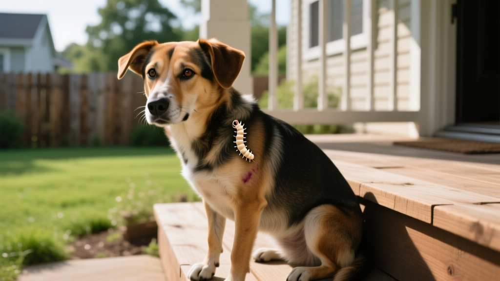
Yes. Dogs can become accidental hosts for Cuterebra botfly larvae — commonly called "wolves" or "warbles" in North America. The larvae typically embed under the skin near the head, neck, or torso, forming a visible swelling with a small breathing hole (fistula). While uncomfortable, most cases are treatable when a veterinarian surgically removes the intact larva. Never attempt to squeeze or extract the larva yourself — rupturing it can trigger a severe allergic reaction or secondary infection. If you notice an unexplained lump with a tiny hole on your dog, consult your veterinarian promptly.
If you've ever spotted a strange, raised bump on your dog's skin with what looks like a tiny pore in the center, you may be dealing with a Cuterebra botfly larva — one of the more unsettling parasites in North American veterinary medicine. While the idea of a maggot-like organism living beneath your dog's skin is alarming, understanding the science behind this parasite can help you act calmly and correctly.
This guide breaks down the full biology of the botfly lifecycle, explains exactly how dogs become infected, outlines what the larva does inside the host's body, and gives you a practical action plan if you suspect an infestation.
The Science Behind Cuterebra: How Botfly in Dogs Actually Works
The Parasite: Cuterebra Species
The botflies that affect dogs belong to the genus Cuterebra, a group of large, robust flies native to the Americas. Unlike common houseflies or blowflies, adult Cuterebra flies do not feed — their sole purpose is reproduction. They are sometimes called "rodent botflies" or "rabbit botflies" because their primary hosts are wild rodents (squirrels, chipmunks, mice) and lagomorphs (rabbits, hares).
Dogs — and cats — are accidental or aberrant hosts. The larvae did not evolve to parasitize canines; dogs simply wander into the wrong place at the wrong time.
The Full Lifecycle: From Egg to Adult Fly
Understanding the lifecycle is critical because it explains exactly how your dog becomes infected — and why prevention is so difficult.
- Egg Deposition (Day 0): The adult female Cuterebra fly lays clusters of 5–15 eggs near the entrances of rodent burrows, rabbit warrens, or along frequently used animal trails. She does not lay eggs directly on a host. Eggs are dark, elongated, and roughly 1 mm long — nearly invisible to the naked eye.
- Egg Activation (Days 1–7): Eggs remain dormant until triggered by the body heat of a passing animal. Temperature change — not physical contact — is the primary hatching stimulus. When a warm-bodied mammal passes within centimeters, the eggs hatch almost instantly, releasing first-stage larvae (L1).
- Larval Entry (Minutes after hatching): The L1 larvae are mobile and seek out a body opening — nostrils, mouth, ears, eyes, or open wounds. In dogs, the most common entry route is through the nose while sniffing at burrow entrances. The larvae attach to mucous membranes and begin migrating.
- Internal Migration (Days 1–10): Depending on the Cuterebra species, the larva migrates through different tissue pathways. Some species travel through the trachea and into subcutaneous tissue. Others migrate through the nasal passages, pharynx, or even the cribriform plate toward the brain (particularly Cuterebra species associated with cerebral myiasis). This migration phase is where neurological complications can arise.
- Warble Formation (Days 7–21): The larva settles in subcutaneous tissue and begins to grow rapidly. It creates a fistula — a small breathing pore connecting its posterior spiracles (breathing organs) to the outside air. The host's body forms a capsule of connective tissue and inflammatory cells around the larva, creating the characteristic "warble" or raised swelling.
- Development and Emergence (Days 21–50+): The larva molts from L1 to L2 and then to L3, growing from roughly 1 mm to 25–40 mm in length. The mature L3 larva exits through the fistula, drops to the ground, burrows into soil, and pupates. The pupal stage lasts weeks to months depending on temperature. An adult fly eventually emerges to restart the cycle.
| Stage | Duration | Location | Size |
|---|---|---|---|
| Egg | 1–7 days (dormant) | Burrow entrances, trails | ~1 mm |
| L1 Larva | Days 1–10 | Migrating through tissue | 1–3 mm |
| L2 Larva | Days 10–25 | Subcutaneous warble | 5–15 mm |
| L3 Larva | Days 25–50+ | Subcutaneous warble | 25–40 mm |
| Pupa | Weeks to months | Soil | ~20 mm |
| Adult Fly | Days to weeks | Free-living (non-feeding) | 15–30 mm |
Evolutionary Context: Why Botflies Target Mammals
An Ancient Parasitic Strategy
The family Oestridae (botflies) has evolved obligate parasitism over tens of millions of years. Fossil evidence and phylogenetic analysis place the origin of botfly-mammal parasitism in the Eocene epoch (~50 million years ago). The strategy is remarkably efficient: rather than spending energy hunting or foraging, the larva co-opts a warm-blooded host as both shelter and food source.
In their natural hosts — wild rodents and rabbits — Cuterebra larvae have co-evolved to cause minimal harm. A heavily parasitized squirrel may carry 2–3 larvae with little impact on survival. This is textbook host-parasite equilibrium: the parasite benefits without killing its host, ensuring the cycle continues.
Why dogs get caught in this cycle: Dogs are curious macrosmatic animals — they investigate the world primarily through their noses. When a dog sniffs at a rodent burrow, its warm breath and body heat trigger egg hatching, and the larvae enter through the nasal mucosa. This is an evolutionary mismatch: the larvae respond to heat and CO₂, not to species-specific cues. Any warm mammal passing by will do.
Physiological Mechanisms: What the Larva Does Inside Your Dog
Immune Evasion and Tissue Manipulation
Once embedded subcutaneously, the Cuterebra larva employs several physiological strategies to survive inside a host whose immune system is actively trying to destroy it:
- Connective tissue capsule manipulation: The larva stimulates the host's body to form a fibrous capsule around it. Rather than fighting this encapsulation, the larva exploits it — the capsule provides structural support and partially shields the larva from circulating immune cells.
- Anti-inflammatory secretions: Research on related Oestridae species has demonstrated that botfly larvae secrete molecules that modulate the host's local immune response, suppressing the inflammatory cascade that would otherwise lead to larval rejection. These include protease inhibitors and immunomodulatory peptides identified in studies published in the National Institutes of Health (NIH) parasitology literature.
- Controlled tissue feeding: The larva feeds on host tissue fluids, exudate, and cellular debris — not on blood vessels or major structures. It grows slowly and methodically, avoiding damage that would trigger a severe systemic response.
- Fistula maintenance: The breathing pore is kept open through mechanical pressure from the larva's posterior spiracles. If the fistula closes, the larva suffocates — which is why occluding the pore with petroleum jelly is sometimes discussed as a removal technique (though this is not recommended for owners; see below).
The Neurological Complication: Cerebral Myiasis
In rare but well-documented cases — particularly in cats and small dogs — Cuterebra larvae migrate aberrantly into the central nervous system. The larva may pass through the cribriform plate (the thin bone separating the nasal cavity from the brain) and enter the brain parenchyma. This condition, called cerebral myiasis, can cause:
- Sudden-onset seizures
- Circling behavior or head tilt
- Blindness (cortical)
- Behavioral changes (aggression, disorientation)
- Paralysis or ataxia
According to the Merck Veterinary Manual, cerebral cuterebriasis is more commonly reported in cats but has been documented in dogs, particularly in the northeastern and southeastern United States. Prognosis depends on the larva's location and the extent of neural tissue damage.
Recognizing Botfly in Dogs: Symptoms and Diagnosis
What to Look For
The most common presentation is a subcutaneous swelling — typically 1–3 cm in diameter — with a small, circular opening (the fistula) at its apex. You may observe:
- A firm, raised lump on the head, neck, chest, abdomen, or near the tail base
- A visible pore or hole in the center of the swelling (2–5 mm diameter)
- Serosanguinous (pinkish) or purulent discharge from the fistula
- Occasional movement visible beneath the skin or within the pore
- Excessive licking, scratching, or biting at the site
- Mild lethargy or reduced appetite (less common)
- Fever in cases with secondary bacterial infection
Seasonal and Geographic Patterns
Botfly cases in dogs follow a distinct seasonal pattern. In temperate North America, infections peak between late July and early October, corresponding to the period when L2 and L3 larvae are developing inside hosts after summer egg-laying. In warmer southern states (Florida, Texas, Georgia), the season may extend from May through November.
Dogs at highest risk are those that:
- Have access to wooded areas, fields, or brushy yards
- Frequently investigate rodent burrows or rabbit holes
- Are sporting, hunting, or herding breeds with high prey drive and nose-to-ground behavior
- Spend unsupervised time outdoors during late summer and early fall
How Your Veterinarian Confirms the Diagnosis
Diagnosis is usually straightforward based on clinical appearance — the warble with its characteristic fistula is nearly pathognomonic. Your veterinarian may:
- Clip hair around the site and examine the fistula with magnification
- Gently probe the opening to visualize the larva's spiracles
- Use ultrasound in ambiguous cases to confirm the presence of a fluid-filled cavity with a moving organism
- In neurological cases, MRI or CT imaging may reveal the larva's migration tract through brain tissue
Typical diagnostic costs range from $50–$150 for a standard exam and warble identification, up to $800–$2,000 for advanced imaging if cerebral involvement is suspected.
Treatment, Removal, and Recovery
⚠️ Critical Warning: Do Not Squeeze the Warble
The single most important piece of advice: never attempt to squeeze, pop, or manually extract the larva at home. If the larva ruptures during removal, its internal contents — including gut bacteria and foreign proteins — flood the surrounding tissue. This can trigger:
- Anaphylactic reaction — rare but potentially fatal
- Severe local inflammation with tissue necrosis
- Secondary bacterial abscess requiring prolonged antibiotic treatment
- Granuloma formation — a chronic inflammatory mass that may require surgical excision
Professional Veterinary Removal
Your veterinarian will typically follow this protocol:
- Sedation or local anesthesia: The area is numbed with lidocaine or the dog is lightly sedated, depending on temperament and warble location.
- Fistula enlargement: The veterinarian carefully widens the breathing pore using a hemostat or scalpel — just enough to allow larval passage.
- Intact extraction: Using fine forceps, the larva is grasped gently and withdrawn whole. The key is slow, steady traction to avoid tearing the larva's cuticle.
- Cavity debridement: The empty capsule is flushed with sterile saline or dilute chlorhexidine to remove debris and reduce infection risk.
- Antibiotics (if needed): A short course of amoxicillin-clavulanate (typically 13.75 mg/kg twice daily for 7 days) may be prescribed if secondary infection is present.
- Wound management: The site is left open to heal by second intention (natural tissue repair) — suturing is generally avoided to allow drainage.
Total cost for straightforward surgical removal typically ranges from $100–$350, depending on your region and whether sedation is required. Complex cases involving neurological migration can cost $2,000–$5,000+ when imaging, neurosurgery, and hospitalization are involved.
Recovery Timeline
Most dogs recover fully within 7–14 days after intact larval removal. The wound contracts and closes gradually. Keep the area clean, prevent licking with an Elizabethan collar if necessary, and monitor for signs of infection (increasing redness, swelling, pus, or fever).
Common Misconceptions About Botfly in Dogs
| Myth | Fact |
|---|---|
| "Botflies lay eggs directly on dogs." | Cuterebra flies lay eggs in the environment — near burrows and trails. They do not land on or touch your dog. The dog picks up larvae by proximity. |
| "You can suffocate the larva with petroleum jelly." | While occluding the fistula can theoretically force the larva to emerge for air, this method is unreliable and risky. The larva may die in place, triggering abscess formation. Veterinary extraction is safer and more effective. |
| "Botfly larvae are the same as maggots from flystrike." | Completely different organisms. Flystrike (myiasis from blowflies) involves surface-feeding maggots on open wounds. Botfly larvae develop inside intact tissue with a defined capsule. |
| "Monthly flea/tick preventives protect against botflies." | Standard parasiticides (isoxazolines like Bravecto, NexGard, Simparica) are not labeled or proven effective against Cuterebra larvae. There is currently no FDA-approved preventive for botfly in dogs. |
| "If the larva comes out on its own, the dog is fine." | Natural emergence can leave an infected cavity, and there's no guarantee the larva exited intact. Always have the site evaluated by a veterinarian even if the warble appears to have resolved. |
| "Only outdoor/rural dogs get botflies." | Suburban dogs are regularly affected — rodent burrows exist in backyards, parks, and greenbelts. Any dog that sniffs at ground level in a vegetated area is at risk during botfly season. |
Practical Prevention: Reducing Your Dog's Risk
Actionable Prevention Strategies
Because no preventive medication exists for Cuterebra, management relies on environmental awareness and behavior modification:
- Inspect your dog daily during peak season (late July–October in most of the US). Run your hands over the head, neck, chest, and abdomen, feeling for small bumps or scabs.
- Restrict access to rodent burrows. Fill or block holes in your yard. Use wire mesh over ground-level openings near foundations.
- Keep grass and brush trimmed. Short vegetation reduces rodent habitat and makes burrow entrances more visible.
- Supervise outdoor time in wooded or field areas. Redirect your dog if it begins intensely sniffing at ground holes.
- Consider a basket muzzle during high-risk outings if your dog is a compulsive burrow-investigator. A Baskerville Ultra or similar basket-style muzzle allows panting and drinking while preventing nose-to-ground contact.
- Perform a full-body check within 2 hours of returning from hikes, hunts, or off-leash field work.
When a "Botfly Bump" Signals Something More Serious
Not every lump on your dog is a botfly warble. Several conditions can mimic the appearance of a Cuterebra infestation:
- Abscess: A bacterial pocket from a bite wound or foreign body (grass awn, thorn). Unlike a warble, abscesses are typically hot, painful, and lack a defined fistula with a visible larva.
- Skin tumor (mast cell tumor, histiocytoma): Can appear as a raised, firm nodule. Mast cell tumors may ulcerate and drain, mimicking a fistula. Any persistent lump should be aspirated by your veterinarian for cytology.
- Foreign body granuloma: A reaction to embedded plant material (foxtails, grass seeds). Common in the ears, between toes, and in the axillary region.
- Injection site reaction: A firm swelling following vaccination, typically at the scruff or lateral thigh. Usually resolves within 2–4 weeks.
🚨 When to Seek Emergency Veterinary Care
Contact your veterinarian or an emergency clinic immediately if your dog shows any of the following alongside a suspected botfly warble:
- Seizures or sudden behavioral changes (possible cerebral migration)
- Difficulty breathing or noisy respiration (larva in nasal passage or trachea)
- Facial swelling beyond the warble site (allergic reaction)
- Eye discharge, squinting, or visible larva near the conjunctiva (ophthalmomyiasis)
- High fever (>103.5°F / 39.7°C) with lethargy
For suspected poisoning or toxic exposure alongside parasitic concerns, contact ASPCA Animal Poison Control: (888) 426-4435 (24/7, consultation fee applies).
How This Differs From Humans and Other Species
While Cuterebra species are primarily New World parasites, humans can occasionally serve as accidental hosts — a condition called furuncular myiasis. However, the human-infecting botfly in Central and South America is a different species: Dermatobia hominis, which uses a phoretic vector (mosquitoes or ticks) to transport its eggs to the host. Cuterebra species that affect North American dogs and cats rarely, if ever, infect humans.
Compared to dogs, cats appear to be more susceptible to the cerebral migration form of cuterebriasis. The reason is not fully understood but may relate to cats' smaller skull size and thinner cribriform plate, making neural penetration easier for a migrating larva.
Frequently Asked Questions
Is a botfly larva in my dog contagious to other pets or people?
No. Cuterebra larvae cannot transfer from one host to another. The larva must complete its development inside the original host or be surgically removed. There is no risk of your other pets or family members "catching" the larva from an infected dog.
Can dogs die from a botfly infestation?
Death from a simple subcutaneous warble is extremely rare. The serious risk comes from aberrant migration — particularly cerebral myiasis — which can cause permanent neurological damage or, in severe cases, death if untreated. Prompt veterinary care virtually eliminates the risk from standard subcutaneous cases.
Will the larva come out on its own if I just wait?
Eventually, a mature L3 larva will exit the warble on its own to pupate — but this can take weeks, during which your dog remains uncomfortable and at risk for secondary infection. Additionally, natural emergence does not guarantee the larva exits intact. Veterinary removal is faster, safer, and more humane.
Are certain breeds more at risk for botfly in dogs?
There is no genetic predisposition to Cuterebra infection. Risk is driven entirely by behavior and environment. Breeds with high prey drive and ground-scenting behaviors — such as Beagles, Dachshunds, Jack Russell Terriers, and various hound breeds — may encounter more rodent burrows and therefore face higher exposure. However, any dog sniffing in the right environment during botfly season is vulnerable.
How much does botfly removal cost at the vet?
For a straightforward subcutaneous warble, expect to pay $100–$350 including exam fee, sedation (if needed), extraction, and any prescribed antibiotics. Complicated cases — such as larvae in the nasal cavity, eye, or brain — require advanced procedures and can cost $1,000–$5,000+ depending on imaging, surgical complexity, and hospitalization needs.
Is there any medication that prevents botfly larvae?
Currently, no FDA-approved product is labeled for Cuterebra prevention in dogs. Some veterinarians have anecdotally suggested that monthly isoxazoline-class preventives (NexGard, Bravecto, Simparica Trio) may reduce larval survival, but this has not been validated in controlled studies. Environmental management remains the primary preventive strategy. Always consult your veterinarian about the best parasite prevention plan for your dog's specific risk profile.
Sources and Further Reading
- Merck Veterinary Manual — Cuterebra Infestation in Small Animals
- NIH / PMC — Immunomodulation by Oestridae Larvae
- AVMA — Parasite Prevention for Pet Owners
This article is for educational purposes only and does not replace professional veterinary advice. Always consult your veterinarian for diagnosis and treatment of parasitic infestations in your dog.
Marcus Aldridge
All our authors care for dogs every day — read more of their work on the authors page.


