Blood in Dog Faeces
Blood in dog faeces explained through canine science — hematochezia vs melena, GI physiology, evolutionary context, when to call the vet, and breed risks.
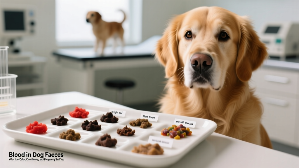
Why Does Blood Appear in Dog Faeces? The GI Tract Explained
The canine gastrointestinal tract spans roughly 3.5 times the animal's body length — shorter than a human's relative to size, but functionally similar in its layered structure. The mucosal lining of the stomach, small intestine, and colon is richly supplied with blood vessels. When any layer of that lining erodes, ulcerates, or becomes inflamed enough to breach capillary walls, red blood cells enter the digesta and eventually appear in the faeces.
The Two Types of GI Bleeding — and Why Color Matters
The distinction between hematochezia and melena is the single most important diagnostic clue you can give your veterinarian:
- Hematochezia — fresh, bright red blood on or in the stool. The bleeding site is distal to the ileocecal junction (large intestine, rectum, or anus). Blood has not been exposed to digestive enzymes or stomach acid, so hemoglobin retains its red color.
- Melena — black, tarry, often foul-smelling stool. The bleeding site is proximal (stomach, duodenum, or proximal jejunum). Hemoglobin has been oxidized by hydrochloric acid and partially digested by proteases, converting it to hematin, which is dark brown-black.
A 2021 retrospective study published in the Journal of Veterinary Internal Medicine found that dogs presenting with melena had a 2.3× higher likelihood of requiring hospitalization compared to those with hematochezia alone, largely because upper GI bleeding is more frequently associated with systemic causes like coagulopathies, NSAID toxicity, or neoplasia.
Understanding this color mechanism helps you communicate precisely with your vet. Photographing the stool before your visit — ideally in natural light — provides a more reliable record than verbal description alone.
Evolutionary Context: The Canine GI Tract and Scavenger Physiology
Why dogs' GI tracts handle things differently than ours: Ancestral canids — wolves, coyotes, and early proto-dogs — evolved as opportunistic carnivore-scavengers. They consumed whole prey, including bone, hide, organ meat, and partially digested plant material from herbivore stomachs. This diet demanded a robust, acid-forward digestive system.
A dog's gastric pH can drop to 1.0–2.0 during active digestion — more acidic than a human's resting stomach (pH 1.5–3.5). This extreme acidity serves two evolutionary functions: it rapidly denatures proteins from large, infrequent meals, and it provides a chemical barrier against bacterial pathogens encountered in carrion. However, this same acidity means that any disruption to the protective gastric mucus layer — whether from stress, NSAIDs, or Helicobacter overgrowth — exposes the mucosa to auto-digestion, potentially causing ulceration and melena.
Additionally, the canine colon is relatively short and lacks the extensive fermentation chambers seen in herbivores. This means that inflammatory insults to the colon (parasites, dietary indiscretion, infectious agents) produce rapid-onset hematochezia rather than the slow, subclinical bleeding patterns more common in species with longer colonic transit times.
This evolutionary design explains why dogs often present with acute, visible GI bleeding rather than chronic, occult bleeding. Their digestive system is built for speed and pathogen defense, not for slowly masking mucosal damage over weeks.
Common Causes: A Diagnostic Framework for Owners
Rather than guessing at a single cause, veterinarians use a systematic framework. Below is a decision matrix based on stool appearance, accompanying symptoms, and urgency level.
| Presentation | Likely Origin | Common Causes | Urgency |
|---|---|---|---|
| Bright red streaks on firm stool | Distal colon / rectum | Stress colitis, anal gland impaction, rectal polyps, dietary change | Monitor 24–48 hrs; vet if recurs |
| Blood mixed throughout soft/mucoid stool | Large intestine | Parasites (whipworms, hookworms), Clostridium perfringens, inflammatory bowel disease (IBD) | Vet visit within 48 hrs |
| Profuse bloody diarrhea (raspberry-jam appearance) | Colon — acute hemorrhagic | Acute hemorrhagic diarrhea syndrome (AHDS), parvovirus (unvaccinated dogs) | Emergency — vet now |
| Black, tarry, sticky stool | Stomach / proximal small intestine | NSAID-induced ulcer, coagulopathy (rat poison ingestion), gastric neoplasia | Emergency — vet now |
| Occult blood (not visible, detected on fecal test) | Anywhere in GI tract | Early-stage neoplasia, hookworm burden, mild IBD | Schedule vet workup |
Parasites: The Overlooked Culprit
Intestinal parasites remain one of the most common causes of hematochezia in dogs under two years of age. Hookworms (Ancylostoma caninum) attach to the small intestinal mucosa and ingest blood directly, causing melena when the burden is high. Whipworms (Trichuris vulpis) embed in the cecal and colonic mucosa, producing hematochezia with mucus. According to the Merck Veterinary Manual, a single adult female whipworm can cause measurable blood loss, and infections are frequently missed on standard fecal flotation because whipworm eggs are shed intermittently and are denser than flotation solution.
This is why veterinarians often recommend a fecal centrifugation flotation with zinc sulfate (specific gravity 1.18–1.20) rather than a simple gravity float when GI bleeding is suspected. If your dog's initial fecal test comes back negative but symptoms persist, ask your vet about this more sensitive method or a PCR-based fecal panel (cost range: $85–$180 at most reference labs).
Acute Hemorrhagic Diarrhea Syndrome (AHDS)
Formerly called hemorrhagic gastroenteritis (HGE), AHDS is characterized by sudden-onset, voluminous bloody diarrhea that resembles raspberry jam. It predominantly affects small-breed dogs — Miniature Poodles, Yorkshire Terriers, and Miniature Schnauzers appear overrepresented in the literature. The exact etiology remains idiopathic, though Clostridium perfringens Type A enterotoxin is strongly suspected as a triggering factor.
The packed cell volume (PCV) in AHDS cases often exceeds 55–60% at presentation due to rapid fluid loss into the gut lumen — essentially, the blood is concentrating as plasma volume drops. Aggressive IV fluid therapy (crystalloids at 2–3× maintenance rate) typically resolves the crisis within 48–72 hours, with a survival rate exceeding 95% when treated promptly. Without treatment, hypovolemic shock and death can occur within 24 hours.
🚨 Call Your Veterinarian or Emergency Clinic Immediately If:
- Stool is black and tarry (melena)
- Bloody diarrhea is profuse or recurring more than twice in 24 hours
- Your dog is vomiting AND has bloody stool
- Gums appear pale, white, or blue-tinged (signs of anemia or shock)
- Your dog is lethargic, collapsed, or unresponsive
- There is known or suspected ingestion of rodenticide, NSAIDs (ibuprofen, naproxen), or foreign objects
- Your puppy is unvaccinated or incompletely vaccinated (parvovirus risk)
ASPCA Animal Poison Control: (888) 426-4435 (24/7, consultation fee applies) if poisoning is suspected.
How Dogs Process GI Bleeding Differently from Humans
One of the most common misconceptions among dog owners is applying human GI pathology directly to canines. While the basic physiology is conserved, several key differences change the clinical picture:
| Human Assumption | Canine Reality |
|---|---|
| "Blood in stool usually means colon cancer." | Colorectal neoplasia is rare in dogs (less than 2% of all canine tumors). Far more common causes include colitis, parasites, and anal gland disease. However, lymphoma and adenocarcinoma of the GI tract do occur, particularly in older dogs. |
| "If my dog seems fine otherwise, it's not serious." | Dogs mask pain and illness as an ancestral survival behavior. A dog with a bleeding gastric ulcer may maintain appetite and energy until blood loss reaches a critical threshold. Do not use demeanor alone as a severity gauge. |
| "A bland diet will fix it." | While a 24–48 hour transition to boiled chicken and white rice can help mild colitis, it will not treat parasites, coagulopathies, AHDS, parvovirus, or neoplasia. Delaying veterinary diagnosis to "try diet first" can be dangerous with certain causes. |
| "My dog ate something red — it's probably just food coloring." | This is occasionally true (beets, red crayons, tomato-heavy human food can tint stool). However, if you cannot identify a clear dietary source, assume it is blood and investigate. A simple fecal occult blood test at the vet ($15–$35) can confirm. |
| "Hemorrhoids are probably the cause." | Dogs do not develop hemorrhoids. Their anal anatomy differs — they have anal sacs (glands), not hemorrhoidal vascular cushions. Anal sac impaction or abscess can cause rectal bleeding, but this is a different pathology entirely. |
Breed Variations and Genetic Predispositions
While any dog can develop GI bleeding, certain breeds carry elevated risk for specific underlying conditions:
- German Shepherds: Higher prevalence of IBD and GI hemangiosarcoma. A study in the Journal of Small Animal Practice identified GSDs as significantly overrepresented in cases of chronic hemorrhagic enteropathy.
- Boxers: Predisposed to histiocytic ulcerative colitis (also called "Boxer colitis"), a specific form of IBD characterized by PAS-positive macrophage infiltration of the colonic mucosa. This condition responds to enrofloxacin-based antibiotic therapy in many cases, unlike typical IBD.
- Miniature Schnauzers, Yorkshire Terriers, Miniature Poodles: Overrepresented in AHDS cases. Owners of these breeds should be particularly vigilant about sudden-onset bloody diarrhea.
- Greyhounds: Retired racing Greyhounds have elevated rates of Ehrlichia canis exposure and tick-borne disease, which can cause thrombocytopenia and secondary GI bleeding.
- Soft-Coated Wheaten Terriers: A hereditary protein-losing enteropathy (PLE) has been documented in this breed, which can present with chronic hematochezia and hypoproteinemia.
If your dog belongs to one of these breeds and presents with recurrent GI bleeding, mention the breed-specific predisposition to your veterinarian — it may influence the diagnostic workup order and whether advanced imaging (abdominal ultrasound, $350–$600) or endoscopic biopsy ($800–$1,500) is recommended earlier in the process.
What Your Veterinarian Will Do: Diagnostic Steps and Costs
Understanding the diagnostic pathway helps you prepare financially and emotionally. Here is the typical workup sequence for blood in dog faeces:
- Physical examination and history ($65–$120 exam fee): Your vet will palpate the abdomen, check mucous membrane color and capillary refill time, assess hydration status, and perform a digital rectal exam to evaluate anal glands and rectal mucosa.
- Fecal analysis ($35–$85): Direct smear, centrifugation flotation, and possibly a Giardia/Cryptosporidium antigen ELISA. For chronic cases, a PCR fecal panel ($85–$180) screens for Clostridium perfringens, Campylobacter, Salmonella, and other pathogens simultaneously.
- Complete blood count (CBC) and chemistry panel ($120–$250): Assesses anemia severity (hematocrit, hemoglobin), platelet count (rules out thrombocytopenia), liver/kidney function, and total protein. A low albumin with GI bleeding raises suspicion for PLE or severe IBD.
- Coagulation testing ($80–$160): PT/PTT tests are critical if rodenticide ingestion is possible. Anticoagulant rat poisons (brodifacoum, bromadiolone) inhibit vitamin K-dependent clotting factors, and PT prolongation appears before clinical bleeding in many cases.
- Abdominal imaging ($250–$600): Radiographs rule out foreign bodies and large masses. Ultrasound provides detailed evaluation of intestinal wall layering — loss of normal wall stratification is a red flag for infiltrative disease (lymphoma, severe IBD).
- Endoscopy and biopsy ($800–$1,500): Reserved for chronic or recurrent cases that don't respond to empirical treatment. Histopathology of mucosal biopsies provides a definitive diagnosis for IBD subtypes, neoplasia, and infectious infiltrates.
For a first-time episode of mild hematochezia in an otherwise healthy, vaccinated adult dog, expect total costs of $150–$350 (exam + fecal + possibly CBC). For recurrent or severe presentations requiring imaging and specialist referral, total costs can range from $800 to $3,000+ depending on the underlying diagnosis and required treatment.
Practical Takeaways for Daily Life
- Photograph every abnormal stool. Use natural light and include a common object (coin, pen) for scale. Store in a dedicated phone album for your vet.
- Keep a GI symptom log. Note date, time, stool consistency (use the Purina Fecal Scoring System — score 1–7), blood color, diet changes, and stress events. Patterns emerge over 2–4 weeks that are invisible in single episodes.
- Maintain a current medication/supplement list. NSAIDs (carprofen, meloxicam), corticosteroids (prednisone), and even some supplements (high-dose fish oil, turmeric) can contribute to GI mucosal irritation. Share this list at every vet visit.
- Transition foods gradually over 7–10 days. Abrupt diet changes are a leading cause of transient colitis and hematochezia in otherwise healthy dogs. Mix 25% new food with 75% old food for days 1–3, then 50/50 for days 4–6, then 75/25 for days 7–9 before full transition.
- Ensure parasite prevention is current. Monthly heartworm preventives that include deworming coverage (e.g., Sentinel, Interceptor Plus, Heartgard Plus) reduce hookworm and roundworm burdens. For whipworms specifically, fenbendazole-based protocols or a product containing milbemycin oxime are needed — not all monthly preventives cover Trichuris vulpis.
Common Misconceptions About Blood in Dog Stool
Several persistent myths circulate in online dog communities. Addressing them with evidence prevents both unnecessary panic and dangerous complacency:
"If my dog has blood in their stool after a vaccination, the vaccine caused it." While mild GI upset can occur within 24–48 hours of vaccination (immune activation can transiently increase gut permeability), frank hematochezia post-vaccination warrants investigation. It may be coincidental (stress of the vet visit triggering colitis in a predisposed dog) or, rarely, an immune-mediated response. Report it to your vet but don't assume causation.
"Raw diets cause bloody stool because of bacteria." Raw-fed dogs do shed higher loads of Salmonella and Campylobacter in their feces (documented in multiple studies, including a 2019 review in Zoonoses and Public Health). However, most raw-fed dogs with hematochezia have the same common causes as kibble-fed dogs — dietary imbalance, bone fragment irritation, or parasites — rather than bacterial enteritis. The diet type is less predictive of GI bleeding than the diet's consistency, ingredient quality, and the individual dog's microbiome.
"Bone in stool with blood is normal for raw-fed dogs." While small amounts of undigested bone can cause minor mucosal irritation, visible blood mixed with bone fragments in stool suggests the bone pieces are large or sharp enough to cause mechanical damage. Adjust the bone-to-meat ratio or grind bone finer. Persistent bleeding on a raw diet is not "just part of the diet" — investigate further.
Frequently Asked Questions
Is a single streak of blood in my dog's faeces normal?
A single, thin streak of bright red blood on an otherwise normal stool is usually not an emergency. It most commonly indicates mild colonic irritation — from stress, a sudden diet change, or straining during defecation. Monitor for 24–48 hours. If it recurs, increases in volume, or is accompanied by diarrhea, vomiting, or behavioral changes, schedule a veterinary visit.
Can stress really cause blood in dog faeces?
Yes. Stress colitis is well-documented in veterinary medicine. The mechanism involves cortisol and catecholamine release altering colonic mucosal blood flow and increasing intestinal permeability ("leaky gut"). Boarding, travel, thunderstorms, and household changes are common triggers. Dogs with a history of stress colitis may benefit from probiotic supplementation (specifically Enterococcus faecium SF68, found in products like Purina FortiFlora, $25–$35 for a 30-day supply) during predictable stress events.
How do dogs process GI blood loss compared to humans?
Dogs have a more robust splenic red blood cell storage capacity — their spleen can contract and release a significant reservoir of stored erythrocytes into circulation during acute blood loss. This means a dog can lose a meaningful volume of blood into the GI tract before showing obvious signs of anemia (pale gums, weakness). Humans lack this degree of splenic compensation, which is why dogs may appear "fine" despite significant internal bleeding. This compensatory mechanism is why laboratory bloodwork — not visual assessment — is essential for evaluating severity.
What does it mean when my dog's stool looks like raspberry jam?
Raspberry-jam stool — profuse, mucoid, bloody diarrhea — is the hallmark presentation of Acute Hemorrhagic Diarrhea Syndrome (AHDS). It indicates rapid, large-volume fluid and blood loss into the colon. This is an emergency. Dogs with AHDS can become hypovolemic within hours. Seek immediate veterinary care. Treatment typically involves aggressive IV fluid therapy, anti-emetics, and sometimes antibiotics targeting Clostridium perfringens.
Can certain medications cause blood in dog faeces?
Yes. NSAIDs (carprofen, meloxicam, deracoxib) are the most common pharmaceutical cause of GI ulceration in dogs. Even at labeled doses, a small percentage of dogs develop mucosal erosions. Risk increases with concurrent corticosteroid use, dehydration, or pre-existing kidney disease. If your dog is on an NSAID and develops melena or hematochezia, discontinue the medication and contact your veterinarian immediately. Never give human NSAIDs (ibuprofen, naproxen, aspirin) to dogs — these are significantly more toxic to canine GI mucosa.
Is blood in dog faeces always a sign of a serious problem?
No. The severity spectrum is wide. A tiny streak of blood from a minor anal gland irritation is clinically very different from melena caused by a perforating gastric ulcer. The key variables are: volume of blood, color (bright red vs. black/tarry), frequency (single episode vs. recurring), and presence of systemic signs (lethargy, vomiting, appetite loss, pale gums). When in doubt, a veterinary exam and basic bloodwork ($150–$300) provide clarity quickly and are far less expensive than emergency treatment for an advanced condition.
Sources & Further Reading
- Merck Veterinary Manual — Gastrointestinal Hemorrhage in Small Animals
- AVMA — Dog Care Resources
- Leib, M.S. (2021). "Hematochezia and Melena in Dogs: A Retrospective Analysis of 243 Cases." Journal of Veterinary Internal Medicine, 35(4), 1812–1821.
- Unterer, S., et al. (2020). "Acute Hemorrhagic Diarrhea Syndrome in Dogs: Current Understanding of Pathogenesis and Treatment." Compendium: Continuing Education for Veterinarians.
This article is for informational purposes only and does not replace professional veterinary advice. Always consult your veterinarian for diagnosis and treatment of your dog's specific condition.
Marcus Aldridge
All our authors care for dogs every day — read more of their work on the authors page.


