ACL Tear in Dogs: The Science of Canine Knee Injuries Explained
Understand the science behind ACL tears in dogs — from cranial cruciate ligament anatomy and evolutionary causes to diagnosis, treatment costs, and recovery.
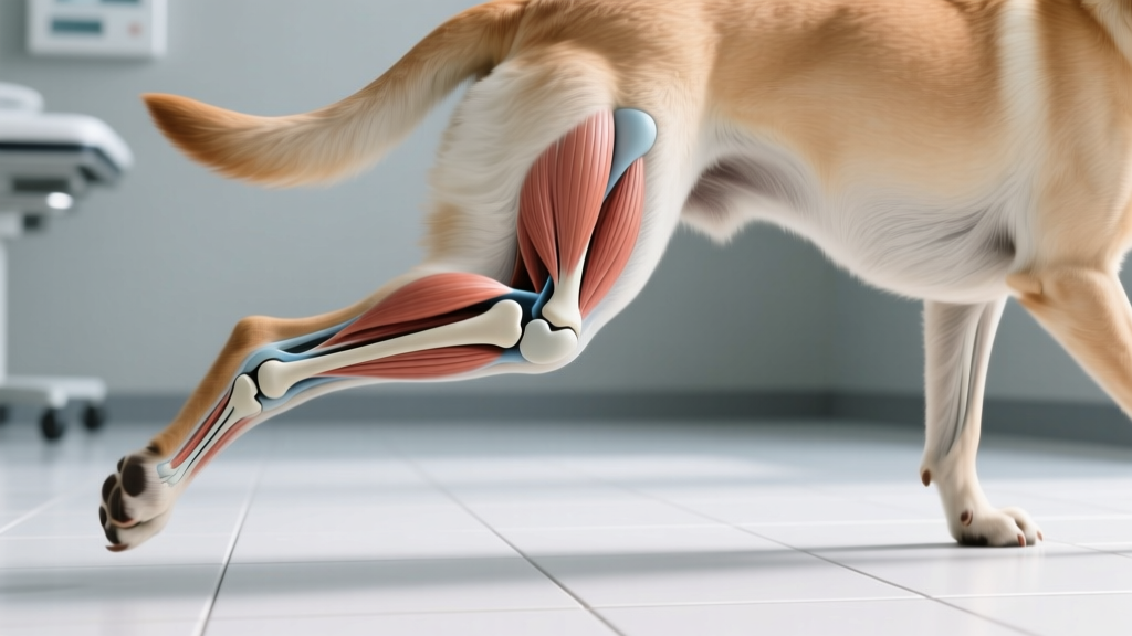
Quick Answer: What Is an ACL Tear in Dogs?
An ACL tear in dogs — properly called a cranial cruciate ligament (CCL) rupture — is a partial or complete tear of the ligament that stabilizes the knee (stifle) joint. Unlike humans who typically tear their ACL during sudden athletic pivots, dogs usually develop CCL tears through gradual degeneration over months or years. The result is hind-leg lameness, joint instability, and progressive arthritis. Treatment ranges from surgical repair ($2,000–$6,000 per knee) to conservative management for dogs under 30 lb. Always consult your veterinarian for diagnosis and treatment planning.
The Anatomy and Physiology Behind CCL Tears
How the Canine Stifle Joint Works
The canine knee — called the stifle — is mechanically similar to the human knee but operates at a fundamentally different angle. Dogs stand with their stifle permanently flexed at approximately 135 degrees, compared to a human's near-straight standing leg. This constant flexion creates a persistent forward-directed force called cranial tibial thrust, which the CCL must resist every single time your dog bears weight.
The CCL is not a single band of tissue. It consists of two functional bundles:
- Cranial band: Taut in both flexion and extension — this is the primary stabilizer and the bundle that most commonly ruptures first
- Caudal band: Taut primarily in extension — provides secondary rotational stability
The ligament also prevents internal rotation of the tibia and limits hyperextension of the stifle. When it tears, the tibia slides forward relative to the femur with each step — a motion veterinarians detect as the cranial drawer sign or tibial compression test.
According to the Merck Veterinary Manual, CCL disease is the single most common orthopedic condition in dogs, affecting an estimated 1 in 50 dogs annually in the United States, with over 600,000 CCL surgeries performed per year.
The physiological mechanism of injury differs sharply from human ACL tears. In humans, roughly 70% of ACL tears are acute, non-contact injuries caused by sudden deceleration, pivoting, or landing awkwardly. In dogs, research published in the Journal of the American Veterinary Medical Association has established that the majority of CCL tears result from chronic, progressive degeneration of the ligament fibers — not a single traumatic event.
Here is what happens at the cellular level:
- Collagen fiber fatigue: Repetitive micro-damage accumulates in the ligament's Type I collagen matrix over months to years
- Mucoid degeneration: The ligament substance undergoes a pathological change where normal dense collagen is replaced by weaker, disorganized mucoid (gel-like) tissue
- Partial tearing: Individual fiber bundles begin to fail, often the cranial band first — this causes intermittent lameness that may come and go
- Complete rupture: The remaining fibers can no longer withstand cranial tibial thrust, and the ligament fails completely, often during a routine activity like jumping off a couch
- Secondary cascade: Joint instability triggers synovial inflammation, meniscal damage (medial meniscus tears occur in 50–70% of complete ruptures), and irreversible osteoarthritis begins within weeks
Evolutionary Context: Why Dogs Are Prone to CCL Tears
From Wolf Anatomy to Modern Dog Knees
The susceptibility of domestic dogs to CCL tears is, in part, an unintended consequence of selective breeding layered on top of ancestral wolf anatomy.
Ancestral wolf locomotion involved long-distance trotting and pursuit over uneven terrain. Wolves have a relatively straight hind-limb conformation with a stifle angle that distributes load efficiently across the joint during sustained movement. Their body mass-to-ligament ratio is well-matched, and their activity patterns involve gradual warm-up and consistent, rhythmic loading.
Domestication changed the equation in several ways:
- Size divergence: Selective breeding created dogs ranging from 2 lb to over 200 lb — far beyond the wolf's 60–120 lb range. Larger breeds carry proportionally greater forces on their stifles, but ligament cross-sectional area did not scale up proportionally
- Conformational extremes: Breeds with exaggerated angulation (German Shepherds with extreme rear angulation) or straight stifles (some Mastiff-type breeds) place abnormal shear forces on the CCL
- Sedentary lifestyle mismatch: A wolf's ligament is conditioned by daily travel of 10–30 miles. A modern dog's weekend warrior pattern — sedentary weekdays followed by intense play — creates the exact fatigue-loading scenario that predisposes to degeneration
- Obesity epidemic: An estimated 56% of pet dogs in the US are overweight or obese (Association for Pet Obesity Prevention, 2022). Each additional pound of body weight multiplies stifle joint forces by 2–4 times during activity
In short, the canine CCL did not evolve to handle the body shapes, weight ranges, and activity patterns that modern breeding and ownership have created. The wolf's knee was optimized for its ecological niche; many modern dogs are operating outside theirs.
Breed Variations and Risk Factors
Not all dogs face equal CCL tear risk. Research has identified clear breed predispositions, along with modifiable and non-modifiable risk factors.
| Category | High-Risk Breeds / Factors | Key Data |
|---|---|---|
| Breed predisposition | Rottweiler, Newfoundland, Labrador Retriever, Staffordshire Terrier, Mastiff-type breeds | Newfoundlands have a 22% lifetime incidence — the highest of any studied breed |
| Size category | Large and giant breeds (>50 lb / 23 kg) | Account for approximately 65% of all CCL ruptures |
| Age at presentation | Peak incidence at 7–10 years | Large breeds present earlier (5–7 years); small breeds later (10–12 years) |
| Body condition | Overweight/obese dogs (BCS ≥ 6/9) | Obese dogs are 2–4x more likely to rupture a CCL |
| Spay/neuter status | Early-spayed/neutered large breeds | Some studies show increased risk when altered before skeletal maturity; evidence remains debated |
| Bilateral involvement | All breeds after first CCL tear | 40–60% of dogs will tear the opposite CCL within 1–2 years |
Diagnosis: How Veterinarians Detect a CCL Tear
If your dog presents with hind-leg lameness, your veterinarian will use a combination of physical examination techniques and imaging to confirm or rule out a CCL rupture. Understanding this process helps you know what to expect.
The Physical Exam
The gold-standard diagnostic tests are:
- Cranial drawer test: The vet stabilizes the femur and attempts to slide the tibia forward. In a CCL-intact stifle, there is minimal movement (< 1–2 mm). A torn CCL allows obvious forward translation. This test requires the dog to be relaxed — tense muscles can mask instability.
- Tibial compression test: The vet holds the stifle at a fixed angle and flexes the hock (ankle). If the CCL is torn, the tibia thrusts forward. This mimics the weight-bearing forces of walking and is often more reliable in anxious or tense dogs.
- Medial buttress palpation: Chronic CCL tears develop a firm thickening on the inside of the knee — a fibrous response to instability. Feeling this "buttress" is a strong indicator of a long-standing partial or complete tear.
Imaging and Diagnostics
Radiographs (X-rays) do not show the ligament itself, but they reveal secondary signs:
- Joint effusion (fluid swelling within the stifle)
- Osteophyte formation (bone spurs) on the tibial plateau and femoral condyles
- Soft tissue swelling patterns consistent with chronic instability
Advanced imaging — MRI or CT — can directly visualize the ligament and is used in ambiguous cases, particularly partial tears where physical exam findings are equivocal. Cost ranges for diagnostic workup:
Typical Diagnostic Costs (2026 Estimates)
- Orthopedic exam: $75–$200
- Stifle radiographs (2–3 views): $200–$450
- Sedation for exam/imaging: $80–$200
- MRI of stifle: $1,500–$3,000 (specialty centers)
- Arthroscopy (diagnostic + therapeutic): $1,500–$3,500
Treatment Options: Surgery vs. Conservative Management
Treatment decisions depend on your dog's weight, age, activity level, comorbidities, and your budget. There is no universally "best" option — each approach has specific indications and trade-offs.
Surgical Options
| Procedure | Mechanism | Best For | Cost Range | Recovery Time |
|---|---|---|---|---|
| TPLO (Tibial Plateau Leveling Osteotomy) | Cuts and rotates the tibial plateau to eliminate cranial tibial thrust; stabilized with a plate and screws | Large/active dogs >50 lb; gold standard for athletic dogs | $3,500–$6,000 | 8–12 weeks restricted; full recovery 4–6 months |
| TTA (Tibial Tuberosity Advancement) | Advances the tibial tuberosity forward to neutralize shear forces via the patellar tendon angle | Medium to large dogs; alternative to TPLO | $3,000–$5,500 | 8–12 weeks restricted; full recovery 3–5 months |
| Lateral Suture (Extracapsular Repair) | Places a heavy nylon suture outside the joint to mimic CCL function; scar tissue eventually stabilizes | Small dogs <30 lb; low-activity dogs; budget-conscious owners | $1,500–$3,000 | 8 weeks restricted; suture may stretch/loosen in large dogs |
| Tightrope CCL (FiberTape) | Uses high-strength suture material (FiberWire/FiberTape) anchored through bone tunnels for extracapsular stabilization | Medium dogs; alternative to TPLO in select cases | $2,500–$4,500 | 8–12 weeks restricted |
For dogs over 50 lb (23 kg), veterinary orthopedic surgeons overwhelmingly recommend TPLO or TTA. The lateral suture technique has a failure rate of 30–50% in large dogs due to suture stretching under load, whereas TPLO success rates exceed 90–95% for return to normal or near-normal function according to the American Veterinary Medical Association resources on orthopedic care.
Conservative (Non-Surgical) Management
Conservative management may be appropriate for:
- Dogs under 30 lb (14 kg) — smaller body mass means less cranial tibial thrust, and fibrous stabilization can develop over 8–12 weeks
- Dogs with significant comorbidities that make anesthesia risky
- Owners with financial constraints (surgery costs can be prohibitive without pet insurance)
A conservative management protocol typically involves:
- Strict rest for 8–12 weeks — leash walks only, no running, jumping, or stairs
- Weight management — target a body condition score (BCS) of 4–5/9; even a 5% body weight reduction significantly decreases stifle loading
- NSAID therapy — carprofen, meloxicam, or grapiprant as prescribed by your vet to manage inflammation and pain
- Physical rehabilitation — underwater treadmill, therapeutic laser, and controlled range-of-motion exercises at a certified canine rehabilitation facility ($75–$150 per session)
- Joint supplements — omega-3 fatty acids (EPA/DHA at 300 mg combined per 10 lb body weight), glucosamine/chondroitin, and green-lipped mussel extract for cartilage support
Be aware: conservative management does not restore joint stability. Osteoarthritis will progress regardless, and approximately 10–15% of small dogs managed conservatively will eventually require surgery due to worsening lameness or meniscal tears.
Common Misconceptions About ACL Tears in Dogs
| Myth | Fact |
|---|---|
| "My dog tore his ACL playing fetch — it was a freak accident." | While acute tears do occur, most canine CCL tears involve pre-existing degeneration. The fetch session was the final straw on a weakened ligament, not the sole cause. |
| "He's limping but still using the leg, so it can't be that serious." | Partial tears cause intermittent lameness that worsens over weeks to months. Early intervention — before complete rupture and meniscal damage — leads to better outcomes. |
| "Surgery fixes the problem completely." | No surgery restores a normal CCL. TPLO and TTA change joint biomechanics to compensate for the missing ligament. Some degree of arthritis is inevitable regardless of treatment. |
| "Small dogs don't need surgery for CCL tears." | Small dogs have better outcomes with conservative management, but many still benefit from surgery — especially if a meniscal tear is present or conservative management fails after 8–12 weeks. |
| "If one knee is torn, the other is fine." | 40–60% of dogs will rupture the contralateral CCL within 1–2 years. The same degenerative process is typically affecting both ligaments simultaneously, and increased load on the "good" leg accelerates its breakdown. |
Practical Takeaways: What This Means for Your Dog's Daily Life
Prevention Strategies Backed by Science
- Maintain lean body condition: Keep your dog at a BCS of 4–5/9. For a Labrador Retriever, that typically means 55–70 lb (25–32 kg) — not the 80+ lb many owners consider "normal." Use a kitchen scale to measure food portions rather than free-feeding.
- Condition before activity: The weekend warrior pattern is a CCL risk multiplier. Aim for consistent daily exercise — 30–60 minutes of moderate activity — rather than weekday inactivity followed by intense weekend hikes.
- Avoid high-impact activities on hard surfaces: Repetitive jumping onto/off concrete, tile, or hardwood floors creates peak stifle loading. Use ramps or stairs for vehicle entry and raised beds.
- Consider pet insurance early: CCL surgery is one of the most common claims. Policies purchased before any lameness diagnosis will typically cover CCL surgery — but most have a 12-month waiting period for orthopedic conditions and may require an orthopedic exam to rule out pre-existing conditions. Premiums range from $30–$80/month depending on breed, age, and location.
- Watch for early warning signs: Intermittent "toe-touching" lameness after exercise, stiffness when rising, reluctance to jump, or a "bunny-hopping" gait are all early indicators of partial CCL degeneration. Early veterinary evaluation can allow treatment before complete rupture.
Post-Surgery Recovery: What to Expect
If your dog undergoes TPLO or TTA, the recovery timeline looks approximately like this:
- Weeks 1–2: Strict confinement, incision monitoring, pain management. Leash-assisted bathroom breaks only. E-collar required.
- Weeks 3–4: Short controlled leash walks (5 minutes, 3–4x daily). Begin passive range-of-motion exercises if recommended.
- Weeks 5–6: Gradual increase in walk duration (10–15 minutes). Recheck radiographs to confirm bone healing.
- Weeks 7–8: If bone healing confirmed, begin off-leash activity in a small, controlled area. Underwater treadmill sessions may start.
- Weeks 9–12: Progressive return to normal activity. Most dogs are using the leg well by week 10–12.
- Months 4–6: Full return to unrestricted activity. Some mild arthritis management may be ongoing long-term.
When to See the Vet: Red Flags for Stifle Problems
Seek Veterinary Evaluation If Your Dog Shows:
- Sudden non-weight-bearing lameness on a hind leg (toe-touching or leg held up entirely)
- Audible "pop" or "snap" during activity followed by lameness
- Progressive stiffness when rising from rest, especially after exercise
- Swelling or warmth around the knee joint
- Reluctance to jump onto furniture, into vehicles, or climb stairs
- Intermittent lameness that improves with rest but returns with activity — this pattern strongly suggests a partial CCL tear in progress
- Sitting with one hind leg extended to the side rather than tucked underneath
Emergency: If your dog is completely unable to bear weight, is vocalizing in pain, or the limb appears deformed or swollen rapidly, seek emergency veterinary care immediately. Acute complete ruptures with meniscal entrapment can cause severe pain.
Frequently Asked Questions
Why do dogs get ACL tears so commonly compared to other animals?
Dogs' permanently flexed stifle angle creates constant cranial tibial thrust — a force that the CCL must resist at all times during weight-bearing. Combined with breed-related conformational extremes, the obesity epidemic in pet dogs, and the mismatch between ancestral ligament conditioning and modern sedentary-then-active lifestyles, dogs face a unique convergence of risk factors. Cats, by contrast, rarely tear their CCL because their lighter body mass and more flexible joints produce far lower shear forces.
Can dogs recover from a CCL tear without surgery?
Yes — but with caveats. Dogs under 30 lb (14 kg) have a reasonable chance of developing sufficient fibrous scar tissue to stabilize the joint within 8–12 weeks of strict rest. Dogs over 50 lb (23 kg) almost never achieve adequate stability without surgery, and prolonged instability accelerates arthritis and increases the risk of a painful meniscal tear. Your veterinarian will help determine which path is appropriate based on your dog's specific situation.
How do dogs sense or show that their CCL is tearing?
Dogs cannot localize pain the way humans do, so they communicate through gait changes rather than pointing to a specific spot. A dog with a degenerating CCL will typically show a "sit test" abnormality — sitting with the affected leg kicked out to the side to avoid stifle flexion. During movement, you may notice a head bob (head rises when the affected leg bears weight), shortened stride on the affected side, and reduced willingness to perform activities requiring stifle flexion like climbing stairs.
What does it mean when my dog is limping but still playing?
Adrenaline and endorphins can mask pain during exciting activities. A dog that limps after a walk but "runs it off" during play is not pain-free — they are overriding pain signals through excitement. This pattern is a classic indicator of early-stage partial CCL degeneration. Continuing high-impact activity on a partially torn ligament accelerates the progression to complete rupture.
Is a CCL tear the same as a luxating patella?
No. These are distinct conditions that affect the stifle but involve different structures. A luxating patella is a kneecap that pops out of its groove, causing intermittent skipping or "catching" of the hind leg. A CCL tear involves the internal stabilizing ligament. However, chronic patellar luxation can increase CCL stress and contribute to ligament degeneration, and the two conditions can coexist — particularly in small breeds.
How is a dog's CCL different from a human's ACL?
They are anatomically homologous — the same structure in a differently oriented joint. The key differences are biomechanical: humans stand with near-straight knees, so the ACL is primarily stressed during athletic pivoting and deceleration. Dogs stand with flexed stifles, so their CCL is under constant load during every step. This fundamental difference explains why human ACL tears are usually acute athletic injuries while canine CCL tears are usually chronic degenerative conditions.
How can I tell the difference between a CCL tear and a hip problem?
Both cause hind-leg lameness, but the patterns differ. CCL tears cause lameness that is typically worse at the start of exercise and may improve slightly as the dog warms up, then worsens again after rest. Hip dysplasia or arthritis tends to cause a more consistent, swaying gait with reduced range of motion in the hip and a "bunny-hopping" rear gait at speed. Your veterinarian can differentiate these through specific orthopedic exam maneuvers and radiographs of both the stifle and hips.
Sources & References
- Merck Veterinary Manual: Cranial Cruciate Ligament Disease in Dogs
- AVMA: Dog Care Resources
- Association for Pet Obesity Prevention
This article is for educational purposes only and does not replace professional veterinary advice. Always consult your veterinarian for diagnosis and treatment of your dog's specific condition.
Anouk Beaumont
All our authors care for dogs every day — read more of their work on the authors page.


